
Fig. 2
|
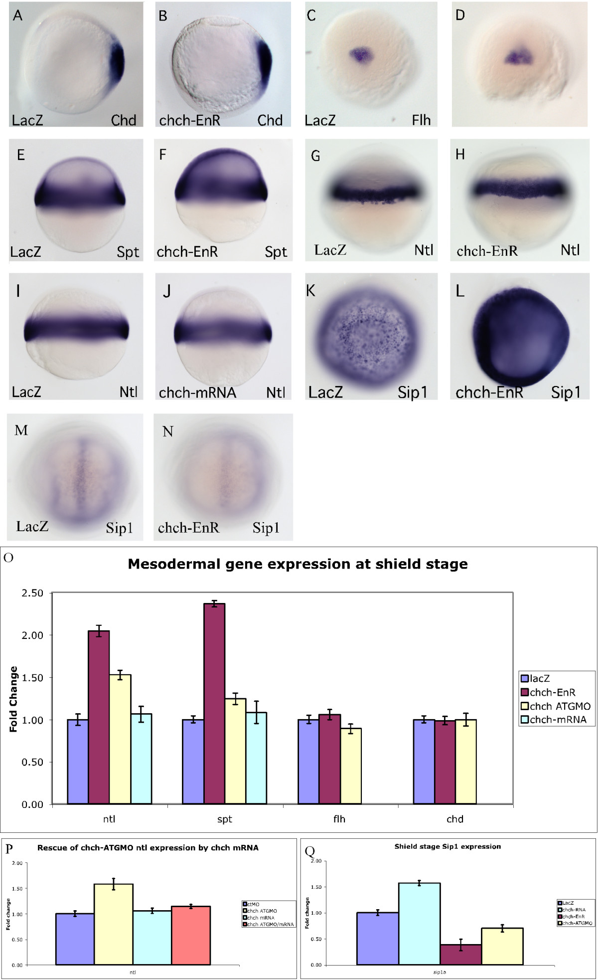
Fig. 2
chch represses mesodermal markers. Mesodermal markers were examined by RNA in situ hybridization and real-time PCR in embryos microinjected with chch-EnR mRNA. Inhibition of chch does not alter expression of dorsal mesodermal genes chd (A, B) or flh (C, D). Expression of mesodermal markers at shield stage, including spt (E-F) and ntl (G-H) are expanded slightly in chch-inhibited embryos. Overexpression of chch mRNA does not alter no-tail expression at shield stage (I-J). chch inhibition results in a decrease in Sip1 gene expression at bud stage (M-N), while chch activation results in Sip1 induction at shield stage (K-L). Real-time PCR analysis of mesodermal markers in chch-inhibited embryos (O). The fold change in transcript levels (y-axis) is graphed relative to control embryos following overexpression of chch-EnR mRNA, chch-ATGMO and chch mRNA. This analysiinhibited embryos (O). The fold change in transcript levels (y-axis) is graphed relative to control embryos following overexpression of