
Fig. 5
- ID
- ZDB-FIG-230728-22
- Publication
- Lin et al., 2023 - YULINK regulates vascular formation in zebrafish and HUVECs
- Other Figures
- All Figure Page
- Back to All Figure Page
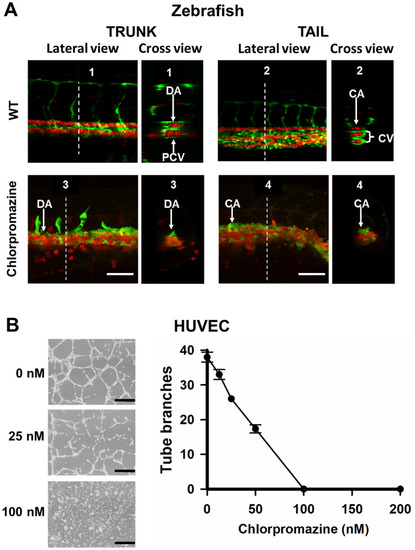
Effects of an endocytosis inhibitor on vein morphogenesis in vivo and capillary tube formation in vitro. A The double Tg (fli1:EGFP; gata1:DsRed) embryos were treated with endocytosis inhibitor, chlorpromazine (60 nM), at 16 hpf. Chlorpromazine was removed at 22 hpf. The lateral and cross-section views of blood vessels in trunk and tail of the Tg (fli1:EGFP; gata1:DsRed) embryos at 24 hpf were shown. White dotted lines indicate the position where the cross-sections are taken. The upper panels were shown as WT groups. DA dorsal aorta, PCV posterior cardinal vein, CA caudal artery, CV caudal vein. Scale bars indicate 50 μm. B Chlorpromazine-mediated inhibition of capillary tube formation in vitro. HUVECs were pretreated with different concentrations of chlorpromazine for 1 h and then seeded in 12-well plates coated with Matrigel. Representative areas are photographed at 10× magnification. Black scale bars indicate 400 μm. Data points represent mean ± SD of two replicate experiments with 4 measurements per treatment |
| Fish: | |
|---|---|
| Condition: | |
| Observed In: | |
| Stage: | Prim-5 |