
Fig. 2
- ID
- ZDB-FIG-230728-19
- Publication
- Lin et al., 2023 - YULINK regulates vascular formation in zebrafish and HUVECs
- Other Figures
- All Figure Page
- Back to All Figure Page
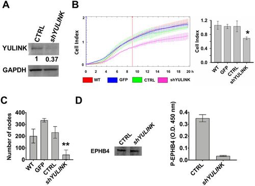
Effects of YULINK-knockdown on formation of capillary-like tubes, migration, and level of phosphorylated EPHB4 in HUVECs. HUVECs were transfected with plasmids containing GFP, vector-control (CTRL), or YULINK shRNA (shYULINK) for 1 day. A The knockdown efficiency of YULINK shRNA was analyzed at 1 day after transfection in Western blot using anti- YULINK antibody. GAPDH was used as a loading control. The intensity of the YULINK band was quantified and normalized to the GAPDH. B Real-time cell migration of the transfected cells was measured using CIM plates in the xCELLigence DP system, which detects the impedance across a cell-permeable membrane. HUVECs were transfected with plasmids containing GFP, vector-control (CTRL), or YULINK shRNA (shYULINK). All cells were seeded (1.6 × 10⁴ cells/well) and allowed to migrate for 20 h (left panel). Cell migration activity, expressed as cell index, was determined 9 h later (right panel, mean ± SD, n = 3, *p < 0.05). WT, wild type untreated cells. C Number of nodes were measured in transfected cells determined using a Matrigel-embedded tube forming assay (mean ± SD, n = 3, **p < 0.001). D Expression of the venous marker EPHB4 in total cell lysates was examined by Western blot and served as the internal control (left panel). Expression of phosphorylation EPHB4 was examined by ELISA (right panel). The ELISA optical density (O.D.) was determined at the visible wavelength 450 nm (mean ± SD, n = 6, **p < 0.001) |