
Fig. 8
- ID
- ZDB-FIG-221226-311
- Publication
- Saettele et al., 2022 - Prolonged Dexamethasone Exposure Enhances Zebrafish Lateral-Line Regeneration But Disrupts Mitochondrial Homeostasis and Hair Cell Function
- Other Figures
- All Figure Page
- Back to All Figure Page
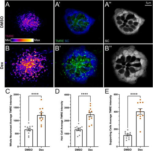
Dexamethasone exposure elevated mitochondrial activity in hair cells and supporting cells. A, B Confocal images of intact pLL neuromasts in live 7 dpf Tg (tnks1bp1:EGFP) fish following TMRE exposure. Fish were treated with DMSO (A) or dexamethasone (B) for 48 h prior to TMRE exposure and imaging. TMRE (A, A′, B, B′) is shown as a max intensity projection and supporting cell (SC) (A′, A″, B′, B″) images are image z-slices from the midlevel of the neuromast. Negative spaces inside GFP-labeled supporting cells indicate neuromast hair cells. LUT bar (A) shows range of intensity values starting at 0 (min) to 11,800 (max). C−E Scatter plots of average TMRE fluorescence intensity (raw integrated density/area values) with SEM bars for whole neuromasts (C), hair cells alone (D), or supporting cells alone (E). Each dot represents the average values of neuromasts L3–L5 for one fish. The data show, compared to DMSO controls, that dexamethasone-treated fish have significantly hyperpolarized mitochondria in both neuromast hair cells and supporting cells (adjusted ****P < 0.0001). N = 1–4 fish per condition per trial; 5 experimental trials |