
Fig. 2
- ID
- ZDB-FIG-221226-306
- Publication
- Saettele et al., 2022 - Prolonged Dexamethasone Exposure Enhances Zebrafish Lateral-Line Regeneration But Disrupts Mitochondrial Homeostasis and Hair Cell Function
- Other Figures
- All Figure Page
- Back to All Figure Page
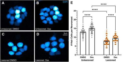
Dexamethasone increased hair cell number in intact and regenerating neuromasts. A–D Representative maximum intensity projection images of 7 dpf neuromasts, with DAPI-labeled hair cell nuclei (blue). Larval lateral-line neuromasts were either left intact (unlesioned) (A, B) or lesioned by treatment with 3 µM CuSO4 for 1 h (C, D). Larvae were then allowed to recover for 2 h, then exposed for 48 h to either 0.1 % DMSO (carrier) (A, C) or dexamethasone (B, D). E Scatter plot graph of hair cell number per neuromast. Bars indicate mean and SEM. Each dot represents a single neuromast on an individual fish. Both intact (unlesioned) and regenerating neuromasts showed a significant increase in the number of hair cells per neuromast in dexamethasone-treated fish relative to DMSO controls (****adjusted P < 0.0001;). N = 5–10 fish per condition per trial; 7 experimental trials |