
Fig. 7
- ID
- ZDB-FIG-221226-305
- Publication
- Saettele et al., 2022 - Prolonged Dexamethasone Exposure Enhances Zebrafish Lateral-Line Regeneration But Disrupts Mitochondrial Homeostasis and Hair Cell Function
- Other Figures
- All Figure Page
- Back to All Figure Page
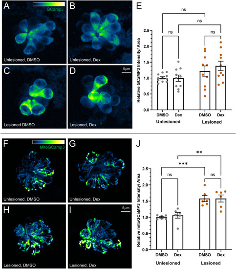
Dexamethasone did not affect hair cell cytosolic or mitochondrial calcium homeostasis. A–D Representative maximum intensity projection images of pLL neuromasts in live 7 dpf Tg(-6myo6b: GCaMP3) fish. The LUT bar in A shows range of intensity values starting at 0 (min) to 2150 (max). E Hair cell cytoGCaMP fluorescence intensity is not affected by dexamethasone exposure. Scatter plots of relative raw integrated density/area values with SEM bars. Each dot represents the average values of neuromasts L3–L5 for one fish. The data show no significant differences in intensity for dexamethasone-treated fish relative to DMSO controls in both intact and regenerating neuromasts. N = 2–3 fish per condition per trial; 5 experimental trials (F–I) Representative maximum intensity projection images of live 7 dpf pLL neuromasts in Tg(-6myo6b: mitoGCaMP3). LUT bar (F) shows range of intensity values starting at 0 (min) to 300 (max). J Hair cell mitoGCaMP fluorescence intensity is not affected by dexamethasone exposure but is elevated in regenerating neuromasts relative to intact neuromasts. Scatter plots of relative raw integrated density/area values with SEM bars. Each dot represents the average values of neuromasts L3–L5 for one fish. The data show no significant differences in intensity for dexamethasone-treated fish relative to DMSO controls in both intact and regenerating neuromasts (adjusted ***P < 0.001 (DMSO), **P < 0.01 (Dex)). N = 2–3 fish per condition per trial; 3 experimental trials |