
Fig. 6
- ID
- ZDB-FIG-221226-310
- Publication
- Saettele et al., 2022 - Prolonged Dexamethasone Exposure Enhances Zebrafish Lateral-Line Regeneration But Disrupts Mitochondrial Homeostasis and Hair Cell Function
- Other Figures
- All Figure Page
- Back to All Figure Page
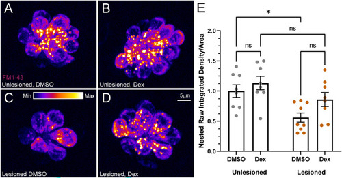
FM1-43 uptake was not significantly affected by dexamethasone exposure. A–D Representative maximum intensity projection images of pLL neuromasts in live 7 dpf immediately after brief (20 s) FM1-43 exposure. Prior to FM1-43 exposure, neuromasts were either left intact (A, B) or lesioned with CuSO4 (C, D), then allowed to regenerate for 48 h while exposed to DMSO (carrier) (A, C) or dexamethasone (B, D). The LUT bar (C) shows the range of intensity values, starting at 0 (min) to 400 (max). E Average FM1-43 intensity per neuromast is not affected by dexamethasone exposure. Scatter plots show raw integrated density (i.e., sum of the pixel values)/area values with SEM bars. Each dot represents the average values of neuromasts L3–L5 for one fish. Values were normalized for each experiment using the inverse median of the unlesioned control values. The data show no significant differences in intensity for dexamethasone-treated fish, relative to DMSO controls (adjusted *P < 0.05). N = 2–3 fish per condition per trial; 4 experimental trials |