
Fig. 3
- ID
- ZDB-FIG-221226-307
- Publication
- Saettele et al., 2022 - Prolonged Dexamethasone Exposure Enhances Zebrafish Lateral-Line Regeneration But Disrupts Mitochondrial Homeostasis and Hair Cell Function
- Other Figures
- All Figure Page
- Back to All Figure Page
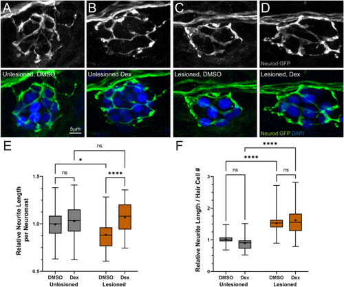
Dexamethasone-enhanced afferent innervation of regenerating neuromasts. A–D Representative maximum intensity projection images of pLL neuromasts in 7 dpf tgBAC (neurod1: GFP) larvae showing afferent neurons (green) and DAPI-labeled hair cell nuclei (blue). Neuromasts were either unlesioned (A, B) or lesioned with CuSO4 (C, D), then allowed to regenerate for 48 h while exposed to DMSO (carrier) (A, C) or dexamethasone (B, D). E, F Box plots of neurite length per neuromast (E) and neurite length per hair cell (F), with whiskers at min/max and mean values marked with “ + ”. Data for each experimental trial were normalized to the median value of unlesioned controls. Regenerating neuromasts (orange plots) in dexamethasone-treated fish had significantly longer neurites per neuromast (E) (****adjusted P < 0.0001); however, the difference in length in dexamethasone-treated fish was not significant relative to the total number of hair cells/neuromast, when compared to DMSO controls (F). N = 4–7 fish per condition per trial; 6 experimental trials |