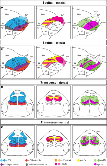
Schematic summary of Figures 1–4. Schemes showing the pattern of marker gene expression and localization of gad1b and vglut2.2-positive areas in the diencephalon of 48 hpf zebrafish. (A,B) Sagittal sections at the medial (A) and lateral (B) levels. (C,D) Transverse section at the dorsal (C) and ventral (D) levels. In the left panel, tcf7l2 expression marks the alar plate of prosomere 1 and 2 and extends into the basal plate; nkx2.2a and lhx9 expression delineate the alar-basal plate boundary in prosomere 1 (CoP, JcP, and PcP) and 2 (Hab, cTh, and rTh); gbx2 and lhx9 expression in the cTh demarcated the rostral border of the Pt; nkx2.2a is also expressed in the rTh, and lhx9 is also expressed in the habenula. In the middle panel, pretectal domains are patterned by the expression of pax6a and pax7a. pax6a is highly expressed in the dorsal subdomain of the Pt. Both pax6a and pax7a markers are present in the central subdomains of the CoP and JcP, but their expression decrease rostrally and ventrally. In the right panel, the expression of gad1b and vglut2.2 identifies several GABAergic and glutamatergic clusters: the Nuc-pc in the caudal edge of the CoP dorsal subdomain, periventricular GABAergic and lateral glutamatergic clusters in the central subdomain of the CoP, lateral GABAergic cluster in the area of the JcP and PcP (lateral parts), and glutamatergic PcP (medial parts). Saturated colors indicate high signal and lighter colors low signal of expression. ap, alar plate; bp, basal plate; Hab, habenula; Mes, mesencephalon; Pt, pretectum; p1, prosomere 1; p2, prosomere 2; p3, prosomere 3; Th, thalamus; cTh, caudal thalamus; rTh, rostral thalamus.
|
