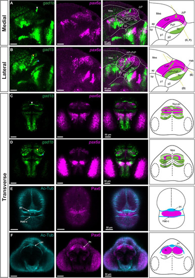
gad1b-positive GABAergic clusters and the identification of Nuc-pc in relation to commissures in the diencephalon of 48 hpf zebrafish. Images showing brain sections stained using immunofluorescence with antibodies specific for acetylated-tubulin and Pax6 (Pax6a and Pax6b), and in situ hybridization with gad1b and pax6a probes. (A,B) In sagittal sections, gad1b and pax6a staining identifies a gad1b-high GABAergic cluster in the dorsal CoP (A, medial section) and two gad1b-high clusters in the central subdomain of the rostral Pt (PcP and JcP) and CoP (B, lateral section). (C) In dorsal sections, co-staining of pax6a and gad1b identifies the gad1b-high cluster as the posterior commissure associated Nuc-pc. (D) In ventral sections, co-staining of pax6a and gad1b identifies a periventricular gad1b-high cluster in the CoP and a lateral gad1b-high cluster in the rostral Pt. (E,F) Acetylated tubulin and Pax6-positive staining identifies the habenular commissure (E) and posterior commissure (F) in transverse sections. White, yellow and blue arrowheads indicate the dorsocaudal, periventricular and lateral gad1b GABAergic clusters, respectively. The schemes depict gad1b-high clusters and the regions identified by the markers in each merged image. Yellow lines show the sectioning plane of the transverse sections. The stripe pattern indicates a co-expression of two markers in a region. ap, alar plate; bp, basal plate; Hab, habenula; Hab-c, habenular commissure; Mes, mesencephalon; Nuc-pc, nucleus of the posterior commissure; pc, posterior commissure; Pt, pretectum; CoP, commissural pretectum; JcP, juxtacommissural pretectum; PcP, precommissural pretectum; p1, prosomere 1; p2, prosomere 2; p3, prosomere 3; Th, thalamus; cTh, caudal thalamus; rTh, rostral thalamus; Pt, pretectum.
|
