FIGURE

FIGURE 5

ID
ZDB-FIG-220402-22
Publication
Brożko et al., 2022 - Genoarchitecture of the Early Postmitotic Pretectum and the Role of Wnt Signaling in Shaping Pretectal Neurochemical Anatomy in Zebrafish
Other Figures
All Figure Page
Back to All Figure Page
FIGURE 5

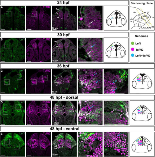
Spatiotemporal expression of the Lef1 and Tcf7l2 proteins in the brain of zebrafish embryos (24–48 hpf). Images showing brain sections from different developmental stages immunostained with antibodies specific for Lef1 and Tcf7l2. (A) 24 hpf:: Tcf7l2-positive cells are detected in the area of the diencephalon mantle zone; lef1 is not expressed in the diencephalon. (B)30 hpf: more Tcf7l2-positive and few Lef1-positive cells are detected in the area of the diencephalon mantle zone. (C) 36 hpf: Lef1-positive cells are visible in the mantle zone in the cTh, caudolateral edge of the Pt and TeO, while Tcf7l2-positive cells are present throughout the Th and Pt with high Tcf7l2 signal in the mantle zone. (D,E) 48 hpf: Lef1-positive cells are visible in the cTh and caudolateral edge of the Pt in the dorsal section (D), being less numerous in the cTh in the ventral section (E), Tcf7l2-positive cells are detected throughout the Th and Pt, being more numerous in ventral sections (E). Yellow lines show the sectioning plane of the sagittal sections in (A–E). Blue arrows point to Tcf7l2-positive cells, yellow arrows point to Lef1-positive cells and white arrows point to double-positive Lef1/Tcf7l2 cells in magnified images. The schemes represent the spatiotemporal distribution of Lef1-positive (green dots), Tcf7l2-positive (magenta dots), and double-positive Lef1/Tcf7l2 (blue dots) cells. Pt, pretectum; TeO, optic tectum; Th, thalamus; cTh, caudal thalamus; rTh, rostral thalamus.

Expression Data

Expression Detail
Antibody Labeling
Phenotype Data

Phenotype Detail
Acknowledgments
This image is the copyrighted work of the attributed author or publisher, and ZFIN has permission only to display this image to its users. Additional permissions should be obtained from the applicable author or publisher of the image. Full text @ Front. Neuroanat.