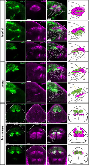
Subdivisions of the pretectum in the brain of 48 hpf zebrafish. Confocal Z-stack images showing brain sections stained using in situ hybridization with dbx1a, pax6a, pax7a, gbx2 and tcf7l2 probes. Sagittal sections: (A)dbx1a expression identifies the PcP in the area of tcf7l2 expression. (B) Co-staining of dbx1a and pax6a shows the alar-basal boundary in prosomere 1 and identifies the pax6a-positive area as the CoP and JcP. (C,D) Co-staining of pax7a and pax6a differentiates between the dorsal, central, and ventral subdomains in the Pt. (E) Staining of pax7a identifies the CoP and JcP and staining of gbx2 identifies cTh in the lateral section. Transverse sections: (F–H)pax7a and gbx2 identify the pretectal domains and the cTh (F), while tcf7l2 stains the Pt and Th (G), and pax6a identifies the CoP and JcP (H). The schemes show the regions identified by the markers in each merged image, with the Pt divided into three rostrocaudal domains: PcP, JcP, and CoP. Stripe pattern indicates co-expression of two markers; saturated color indicates high expression signal while lighter color indicates low signal. The yellow lines (C–E) show the sectioning plane of the transverse sections (F–H). The white arrowhead shows the dbx1a cluster in the dorsal pretectum (B). The white arrow shows the pax7-negative area between the CoP and JcP. ap, alar plate; bp, basal plate; Hab, habenula; Mes, mesencephalon; p1, prosomere 1; p2, prosomere 2; p3, prosomere 3; Pt, pretectum; PcP, precommissural pretectum; JcP, juxtacommissural pretectum; CoP, commissural pretectum; Pth, prethalamus; Th, thalamus; cTh, caudal thalamus; rTh, rostral thalamus; Rh, rhombencephalon.
|
