|
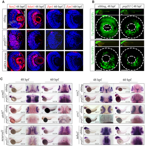
Deletion of Prpf31 impaired RPCs differentiation. (A) Retinal sections of WT, prpf31−/− and prpf31−/− mRNA-rescued embryos were immunostained with Sox2 (marker for RPCs), Islet1 (marker for neuron cells), Zpr1 (marker for cone cells), and Zpr3 (marker for rod cells) antibodies at 48 or 60 hpf. n = 8 for each panel. V, ventral side, D, dorsal side. Scale bar, 100 μm. (B) The distributions of Neurod1:EGFP (specialized neurons, upper panels) and Huc:EGFP (post-mitotic neurons, lower panels) labeled cells in whole-mount retinas from WT and prpf31−/− transgene zebrafish. The dashed circles shown the eyes and lens respectively. n ≥ 7 for each panel; Scale bar, 100 μm. (C) In situ staining of markers for RPCs (ccnd1, vsx2) and for neural precursors (atoh7, crx1), specialized neurons (neurod1) and mature neurons (tuba1) at 48 and 60 hpf. The accumulation of RPCs and reductions of differentiated neurons are shown. Scale bar, 100 μm.
|