|
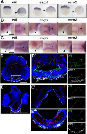
irf6,esrp1 and esrp2 are co-expressed in the oral epithelium of zebrafish embryos. (A-C) Whole-mount in situ hybridization (WISH), showing that irf6, esrp1 and esrp2 maternal deposited transcripts are detected at the eight-cell and shield stage (A; arrowheads indicate periderm), and circumscribe the developing stomodeum and line the oral epithelium of zebrafish embryos at 48 (B) and 72 (C) hpf (arrowheads). All whole-mount embryos are oriented with anterior left and dorsal top. (D-E″) Coronal sections of 48 (D) and 72 (E) hpf embryos analyzed by RNAscope in situ hybridization (ISH), (dorsal top), showing cellular RNA co-expression of irf6 (green) and esrp1 (white) in surface and oral epithelial cells. sox10 (red) staining depicts cartilage elements of the palate. Boxed areas are shown at higher magnification in D′, E′ and E″. Scale bars: 250 μm (A) and 100 μm (B-E″).
|