
Fig. 1
- ID
- ZDB-FIG-200723-46
- Publication
- Xu et al., 2020 - Unifying Developmental Programs for Embryonic and Post-Embryonic Neurogenesis in the Zebrafish Retina
- Other Figures
- All Figure Page
- Back to All Figure Page
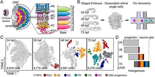
scRNA-seq of embryonic RPCs at different developmental stages. (A) Schematic showing that the zebrafish retina is composed of structurally and functionally radial retinal segments (dashed sector), which undergo the same developmental program in a sequential order (indicated by the arrows: from the center to the periphery). During the developmental program, RPCs give rise to different retinal types in the stereotyped order (indicated on the right). (B) Schematic workflow for scRNA-seq of staged embryonic RPCs using 10x Genomics technology. (C) t-SNE plots show the clustering of qualified retinal cells at different developmental stages (24, 36, 48 and 72 hpf). Different colors represent distinct cell types. The numbers of cells are indicated. (D) Plot showing proportions of distinct retinal cell types (indicated as different colors) over time, from the data in C. AC, amacrine cell; BC, bipolar cell; CMZ, ciliary marginal zone; HC, horizontal cell; MC, Müller glial cell; PR, photoreceptor cell; RGC, retinal ganglion cell. |