
Fig. 2
- ID
- ZDB-FIG-181130-14
- Publication
- Petratou et al., 2018 - A systems biology approach uncovers the core gene regulatory network governing iridophore fate choice from the neural crest
- Other Figures
- All Figure Page
- Back to All Figure Page
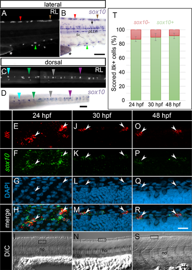
sox10 expression is maintained throughout iridophore development. (A, B) lateral views of the anterior tail of a single embryo at 72 hpf, imaged live under reflected light (RL) (A) and then post-WISH to detect sox10 transcript (B). (C, D) dorsal views of the trunk and anterior tail of a second individual pre- (C) and post- (D) WISH processing for sox10 expression at 72 hpf. Differently coloured arrowheads point to individual iridophores expressing sox10. sox10 is also detected in developing oligodendrocytes (B, arrow), Schwann cells (B) and in iridophores along the yolk sac stripe (A, B asterisk). ltk-positive cells detected via RNAscope (E, J, O arrowheads) all show sox10 transcript (F, K, P; H, M, R arrowheads), at each of 24 (E-I), 30 (J-N) and 48 hpf (O-S). At 24 hpf, cells on the medial migration pathway are shown (I, boxed region). At 30 hpf and at 48 hpf, cells along the developing dorsal stripe are presented (N, S, boxed regions). (T) Quantification of the proportion of ltk+ cells co-expressing (green), or not co-expressing (red) sox10 by RNAscope, at 24, 30 and 48 hpf. Error bars indicate the corresponding standard deviations. (E-S): lateral views of single focal planes. (A-S): heads positioned towards the left. Sc, Schwann cells; pLLn, posterior lateral line nerve; no, notochord; RL, reflected light. Scale bars: (A-D) 100 μm; (E-H, J-M, O-R) 20 μm; (I, N, S) 50 μm. |
| Genes: | |
|---|---|
| Fish: | |
| Anatomical Terms: | |
| Stage Range: | Prim-5 to Protruding-mouth |