
Fig. S11
- ID
- ZDB-FIG-180206-9
- Publication
- Singh et al., 2017 - Different developmental histories of beta-cells generate functional and proliferative heterogeneity during islet growth
- Other Figures
- All Figure Page
- Back to All Figure Page
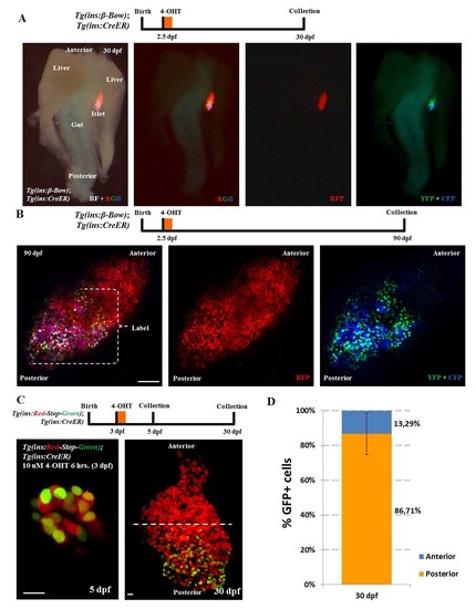
Genetic-tracing of embryonic beta-cells. (A) Whole-mount of the pancreas, liver and the intestine dissected from beta-bow animals at 30 dpf. Embryos were incubated with 4-OHT at 2.5 dpf in order to induce recombination and multicolor labeling of beta-cells. The bright-field (BF) overlaid with fluorescence images shows the localization and orientation of the zebrafish primary islet. The islet lies between the gut and one of the two lobes of the liver. The multicolor beta-cells undergoing recombination at 2.5 dpf (YFP+CFP-positive cells) exhibit predominant localization near the posterior regions of the islet at 30 dpf, whereas the anterior region is composed of cells exhibiting the default red-fluorescence of beta-bow. Anterior is to the top of the images. (B) Maximum intensity projection of islets from 90 dpf (adult) beta-bow animals. Embryos were incubated with 4-OHT at 2.5 dpf. The beta-cells undergoing recombination at 2.5 dpf (YFP+CFP-positive cells) cluster near the posterior regions of the adult islet at 90 dpf, whereas the anterior region contains un-recombined cells, exhibiting the default red-fluorescence of beta-bow. (C) Top – schematic for tracing of embryonic beta-cells during growth. Tg(ins:mCherry-Stop-H2B-EGFP); Tg(ins:CreERT2) animals were incubated with 10 uM 4-OHT at 3 dpf in order to label the embryonic beta-cells with nuclear GFP fluorescence. Samples were collected at 5 and 30 dpf. Bottom - representative maximum intensity projection of primary islets from recombined Tg(ins:mCherry-Stop-H2B-EGFP); Tg(ins:CreERT2) animals at 5 and 30 dpf. Dashed line marks the center of the 30 dpf islet’s anterior-posterior axis. At 5 dpf, 53 ± 12% of the beta-cells were GFP+ (n = 11 islets). (D) Quantification showing the percentage of GFP+ beta-cells present within the anterior and posterior region of the 30 dpf islets. 86 ± 12 % of the GFP+ beta-cells were located in the posterior half of the islet, while 13 ± 12 % of the GFP+ beta-cells were located in the anterior half (n = 7 islets). Error bars = S.D. Scale bar in B, 100 μm; 10 μm in C. |