
Fig. S18
- ID
- ZDB-FIG-180206-14
- Publication
- Singh et al., 2017 - Different developmental histories of beta-cells generate functional and proliferative heterogeneity during islet growth
- Other Figures
- All Figure Page
- Back to All Figure Page
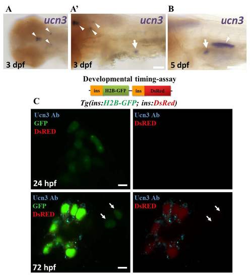
ucn3 expression in zebrafish embryos and larvae. (A-B) Whole mount in-situ hybridization for ucn3 (purple). (A) At 3 dpf, ucn3 transcripts are expressed in characteristic groups of neurons in the brain (arrowheads in A and A’), consistent with a previous report1. In addition, ucn3 is expressed in the islet (arrow in A’). (B) An arrow points to the expression of ucn3 in the islet at 5 dpf. Ucn3 might also show expression in the cells forming the swim-bladder (arrowhead in B). (C) Primary islets from Tg(ins:H2B-GFP; ins:DsRed) embryos at 24 and 72 hpf. EGFP and DsRed are both expressed under the insulin promoter. The relatively faster maturation of GFP compared to DsRED allows to transiently mark the recently-differentiated beta-cells with only green fluorescence and to distinguish them from older beta-cells, which express both colors. At 24 hpf, the GFP-positive cells lack detectable Ucn3-immunofluorescence, whereas at 72 hpf, a majority of the GFP-positive cells are Ucn3-positive. The arrows point to two newborn beta-cells (H2B-GFP-positive/DsRed-negative cells) adjacent to the islet. These cells are Ucn3-negative. The images represent single confocal planes, which were acquired using the same confocal settings at both 24 and 72 hpf. Scale bars in A and B, 1 mm; 5 μm in C. |