
Fig. S20
- ID
- ZDB-FIG-180206-16
- Publication
- Singh et al., 2017 - Different developmental histories of beta-cells generate functional and proliferative heterogeneity during islet growth
- Other Figures
- All Figure Page
- Back to All Figure Page
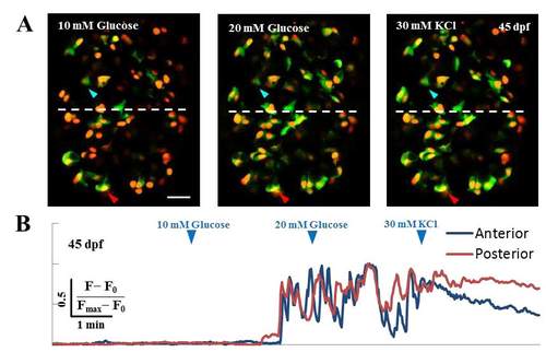
Glucose-stimulated calcium fluxes in beta-cells at 45 dpf. (A-B) Glucose-responsiveness of larval beta-cells at 45 dpf. (A) Islets from Tg(ins:GCaMP6s); Tg(ins:mKO2-nls) animals were mounted ex-vivo and live-imaged while providing increasing concentration of glucose from 5 mM (basal) to 10 and 20 mM, followed by addition of 30 mM KCl. The islets were divided into two halves along the anterior-posterior (A/P) axis, as indicated by the white dotted line (anterior is to the top). Representative cells within each half are marked with arrowheads (anterior – blue; posterior – red). (B) The fluorescence-trace for both cells is shown below in their respective colors. Both cells responded to the stimulation with glucose, as indicated by the oscillating GCaMP6s fluorescence-intensity. Scale bars, 20 μm. |