
FIGURE
Fig. S19
- ID
- ZDB-FIG-180206-15
- Publication
- Singh et al., 2017 - Different developmental histories of beta-cells generate functional and proliferative heterogeneity during islet growth
- Other Figures
- All Figure Page
- Back to All Figure Page
Fig. S19
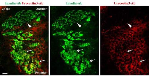
A gradient of Urocortin3 expression along the A/P axis of primary islets at 25 dpf. A single confocal plane of a primary islet from wild-type animals stained using antibodies against insulin and mouse-Urocortin3 (polyclonal) at 25 dpf. The islet is aligned along A/P axis (anterior is to the top). Beta-cells in the posterior region of the islet exhibit positivity for both insulin (green) and Urocortin3 immunofluorescence (red) (arrows). In contrast, the insulin-positive cells in the anterior region are Urocortin3-negative (arrowhead). Scale bars, 20 μm. |
Expression Data
Expression Detail
Antibody Labeling
Phenotype Data
Phenotype Detail
Acknowledgments
This image is the copyrighted work of the attributed author or publisher, and
ZFIN has permission only to display this image to its users.
Additional permissions should be obtained from the applicable author or publisher of the image.
Full text @ Nat. Commun.