
Fig. 3
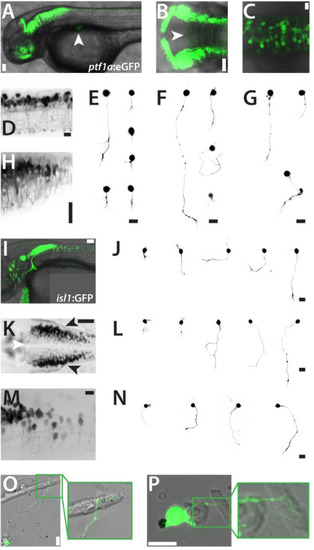
Inhibitory neurons and cranial motoneurons in culture. (A) Tg(ptf1a:eGFP)jh1 embryo (lateral view, 2 dpf). The developing pancreas is marked with a white arrowhead. Scale bar, 50 µm. (B) Hindbrain of a Tg(ptf1a:eGFP)jh1 embryo (dorsal view, 2 dpf) showing eGFP expression in the ventricular zone and long contralateral projections. The white arrowhead indicates the midline. Scale bar, 50 µm. (C,D) Dorsal and lateral details, respectively, of the spinal cord of a Tg(ptf1a:eGFP)jh1 embryo (2 dpf). Scale bars, 50 µm. (H) Lateral detail of the hindbrain/spinal cord transition zone in a Tg(ptf1a:eGFP)jh1 embryo (2 dpf). Scale bar, 150 µm. Anterior is always to the left. (E,F,G)ptf1a:eGFP-positive cells in culture displaying representative morphologies at 1, 2 and 3 dap, respectively. Scale bars, 10 µm. (I) Tg(isl1:GFP)rw0 embryo (lateral view, 2 dpf). Scale bar, 50 µm. (K) Hindbrain of a Tg(isl1:GFP)rw0 embryo (dorsal view, 2 dpf) showing GFP expression cranial motorneurons. The white arrowhead indicates the midline. Scale bar, 50 µm. (M) Lateral detail of the hindbrain/spinal cord transition zone in a Tg(isl1:GFP)rw0 embryo (2 dpf). Scale bar, 50 µm. Anterior is always to the left. (J,L,M)isl1:GFP-positive cells in culture displaying representative morphologies at 1, 2 and 3 dap, respectively. Images were sorted by the size of the cells. Scale bars, 10 µm. (O,P)isl1:GFP positive cells project neurites towards myocytes (2 dap). Scale bars, 10 µm. The figure shows a collection of the images of two preparations for each Tg(ptf1a:eGFP)jh1 and Tg(isl1:GFP)rw0 with comparable results. |
Reprinted from Developmental Biology, 430(1), Sassen, W.A., Lehne, F., Russo, G., Wargenau, S., Dübel, S., Köster, R.W., Embryonic zebrafish primary cell culture for transfection and live cellular and subcellular imaging, 18-31, Copyright (2017) with permission from Elsevier. Full text @ Dev. Biol.