
Fig. 2
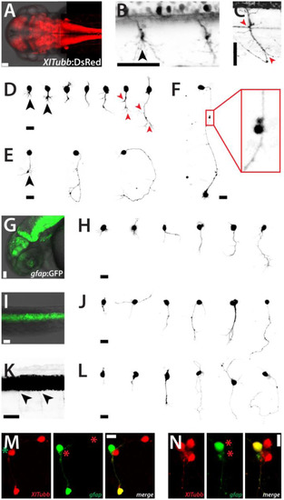
Neurons and glia cells in culture. (A) Head and trunk of a Tg(XITubb:DsRed)zf148 embryo (dorsal view, 2 dpf). Scale bar, 50 µm. (B) Detail of the spinal cord of a Tg(XITubb:DsRed)zf148 embryo (dorsal view, 2 dpf). Motorneurons project highly branched axons (black arrowhead). Scale bar, 50 µm. (C) Detail of the spinal cord of a Tg(XITubb:DsRed)zf148 embryo (lateral view, 2 dpf). A motorneuron with the typical bended axon with several characteristic branching points (red arrowheads) can be observed. Scale bar, 50 µm. Anterior is always to the left. (D,E)XITubb:DsRed-positive neurons in culture displaying representative morphologies at 1 and 2 dap, respectively. Images were sorted by the size of the neurons. Three neurons show highly branched processes like in (B) (black arrowheads). Two neurons strongly resemble the morphology of the motorneuron shown in (C) (red arrowheads). Scale bars, 10 µm. (F) Two XITubb:dsRed-positive neurons at 3 dap forming a putative contact site. Scale bar, 10 µm. (G,I) Head (lateral view) and spinal cord (dorsal view) of a Tg(gfap:GFP)mi2001 embryo (2 dpf). Scale bars, 50 µm. (K) Spinal cord of a Tg(gfap:GFP)mi2001 embryo (lateral view, 2 dpf) displayed at high contrast. Fine cellular protrusions are visible which may represent glia-derived motorneurons still containing stable GFP (black arrowheads). Scale bar, 50 µm. Anterior is always to the left. (H,J,L)gfap:GFP-positive cells in culture displaying representative morphologies at 1, 2 and 3 dap, respectively. Images were sorted by the size of the cells. Scale bars, 10 µm. (M,N) Cultured cells of Tg(XITubb:DsRed)zf148 / Tg(gfap:GFP)mi2001 embryos show overlapping expression of both fluorescent reporters at 2 and 3 dap, respectively. Scale bars, 10 µm. The figure shows a collection of the images of three preparations for Tg(XITubb:DsRed)zf148 and two preparations for Tg(gfap:GFP)mi2001, respectively, with comparable outcomes. |
Reprinted from Developmental Biology, 430(1), Sassen, W.A., Lehne, F., Russo, G., Wargenau, S., Dübel, S., Köster, R.W., Embryonic zebrafish primary cell culture for transfection and live cellular and subcellular imaging, 18-31, Copyright (2017) with permission from Elsevier. Full text @ Dev. Biol.