
Fig. 8
- ID
- ZDB-FIG-160927-16
- Publication
- Robra et al., 2016 - The Intracellular Signaling Molecule Darpp-32 Is a Marker for Principal Neurons in the Cerebellum and Cerebellum-Like Circuits of Zebrafish
- Other Figures
- All Figure Page
- Back to All Figure Page
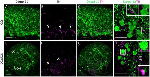
Darpp-32 and TH expression in the adult zebrafish brain. (A-C) TH-positive fibers (open arrow heads in B) run along the Purkinje cell bodies in the CCe. Collapsed z-stacks of confocal images of a coronal section. Scale bar in (A-C) 20 µm. (D) Higher magnification image showing proximity of TH puncta to Purkinje neuron somata (D′, inset size 11.09× 11.09 µm) and dendrites (D′′, inset size 9.98× 9.98 µm). Scale bar in (D) 20 µm. (E-G) Crest cells in the medial octavolateralis nucleus (MON) express Darpp-32. Their dendrites extend into the cerebellar crest (CC), Darpp-32 positive cell bodies are restricted to the MON. TH positive fibers densely innervate the MON. Mild staining in the TH channel in the CC is unspecific staining of blood vessels. Scale bar in (E-G) is 50 µm, sagittal section. (H) Higher magnification image showing TH immunoreactive puncta in close proximity to MON crest cells (H′, inset size 9.98× 9.98 µm) and dendrites (H′′, inset size 4.28× 4.28 µm), coronal section. Scale bar in (H) 20 µm. |
| Genes: | |
|---|---|
| Antibody: | |
| Fish: | |
| Anatomical Terms: | |
| Stage: | Adult |