
Fig. 9
- ID
- ZDB-FIG-160927-17
- Publication
- Robra et al., 2016 - The Intracellular Signaling Molecule Darpp-32 Is a Marker for Principal Neurons in the Cerebellum and Cerebellum-Like Circuits of Zebrafish
- Other Figures
- All Figure Page
- Back to All Figure Page
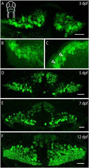
Darpp-32 expression over the course of development. Single optical sections of whole mount immunofluorescence stainings (A,D-F) and projections of a z-stack (B,C). The images are oriented as follows: (A,B,D-F) rostral up, (C) dorsal up, midline to the right. Schematic in (A) is for orientation, the images are showing the cerebellum, indicated by a green box. (A) Darpp-32 expression in the cerebellum is visible at 3 days post-fertilization (dpf). At this stage only cell bodies are visible in the dorsal view. (B) Dorsal view of a 3D projection of the left hemisphere of a 3 dpf larva. (C) Rotation of this 3D-projection around the lateral axis reveals budding dendrites growing towards the dorsal side (white arrowheads). (D-F) Darpp-32 continues to be expressed in the cell bodies and dendrites of Purkinje cells throughout the course of development. In (F) the outgrowing valvula cerebelli can be seen at rostral end of the cerebellum. Scale bar: 20 µm. |
| Gene: | |
|---|---|
| Antibody: | |
| Fish: | |
| Anatomical Terms: | |
| Stage Range: | Protruding-mouth to Days 7-13 |