
FIGURE
Fig. 8
- ID
- ZDB-FIG-151113-17
- Publication
- Kimura et al., 2015 - Integration of vascular Systems between the brain and spinal cord in zebrafish
- Other Figures
- All Figure Page
- Back to All Figure Page
Fig. 8
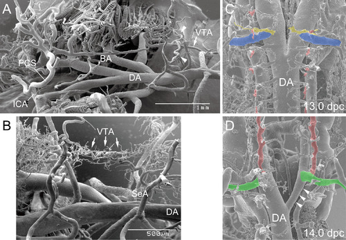
Vascular casts of adult zebrafish and rat embryos. Scanning electron microscopic images of vascular casts of adult zebrafish (dorso-lateral view; A and B) and rat embryos at 13.0 (C) and 14.0 dpc (D; both dorsal view). (C and D) VTA in formation is colored in red; axial vessel for upper limb in blue; seventh SeA in yellow; and subclavian artery in green. Arrows in A and B indicate the VTA. Arrowheads in A indicate the transverse vessel from Se to VTA. Arrowheads in D indicate the regressing aortic arch. |
Expression Data
Expression Detail
Antibody Labeling
Phenotype Data
Phenotype Detail
Acknowledgments
This image is the copyrighted work of the attributed author or publisher, and
ZFIN has permission only to display this image to its users.
Additional permissions should be obtained from the applicable author or publisher of the image.
Reprinted from Developmental Biology, 406(1), Kimura, E., Isogai, S., Hitomi, J., Integration of vascular Systems between the brain and spinal cord in zebrafish, 40-51, Copyright (2015) with permission from Elsevier. Full text @ Dev. Biol.