
Fig. 4
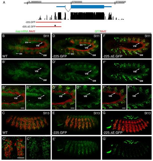
hoip is expressed in striated but not visceral muscle progenitors. (A) hoip gene organization and conservation within the Drosophila genus. The red line identifies genomic sequences used to generate the -225.nGFP and -225ΔE.nGFP hoip reporter genes. (B) St11 embryo labeled for hoip mRNA (green) and Mef2 (red). hoip mRNA is expressed in the Mef2-expressing cells of the somatic mesoderm, as well as in the fat body and the endoderm, but is absent from the neuroectoderm. (B2) hoip expression alone. (B2,B22) High magnification micrograph of the mesoderm shows hoip mRNA expression in the somatic but not the visceral mesoderm. (C-C2) At St13, hoip mRNA is still detectable in the developing somatic musculature. (D-E2) Hoip.-225.GFP embryos labeled for GFP (green) and Mef2 (red). GFP expression recapitulates hoip mRNA expression at St11 (D) and St13 (E). (F-G2) Hoip.-225ΔE.GFP embryos labeled for GFP (green) and Mef2 (red). GFP expression recapitulates hoip mRNA expression at St11 (F) but is undetectable at St13 (G). SM, somatic mesoderm; VM, visceral mesoderm; EN, endoderm. Scale bars: 20 μm. |