
Fig. 1
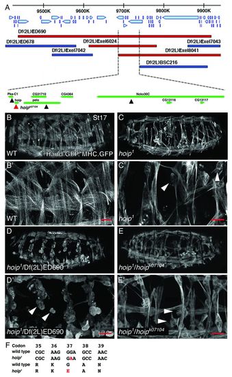
A mutation adversely affecting somatic muscle development maps to hoip. (A) The genomic region uncovered by Df(2L)ED690. Genes and direction of transcription are shown with blue arrows. Deficiencies that fail to complement hoip1 are shown in red; deficiencies that complement hoip1 are shown in dark blue. The minimal overlapping area among the deficiencies that fail to complement hoip1 contains eight genes. Of the four lethal transgene insertions (triangles) in the minimal overlapping area, only P{lacW}hoipk07104 (red triangle) failed to complement hoip1. (B-E) MHC.τGFP, Hand.nGFP expression in St17 embryos. (B) Wild-type embryos express membrane-localized τGFP in each somatic muscle in all embryonic segments. Somatic muscles are severely rounded (arrowheads) in hoip1 (C), hoip1/Df(2L)ED690 (D) and hoip1/P{lacW}hoipk07104 embryos (E). (B2-E2) High-magnification views of embryos shown in B-E. (F) hoip1 is a G37E missense mutation (see supplementary material Fig. S1I). In this and subsequent figures, embryos are oriented with anterior towards the left and dorsal towards the top. Coordinates refer to base pair positions on chromosome 2L. Scale bars: 20 μm. |