
Fig. 2
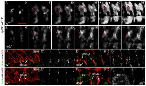
hoip embryos have myotube elongation defects. (A,B) Time-lapse images of rp298>τGFP embryos initiated at late St12. (A) Wild-type embryos showed robust myotube elongation at 30 minutes (double arrows) and developed extensive filopodia for attachment site recognition at 60 minutes (white arrows). (B) Myotubes established polarity in hoip1 embryos at 15 minutes but failed to elongate by 30 minutes. Polarized myofibers at 15 minutes compacted over time (double-headed arrows). (C,D) St16 rp298>τGFP embryos double labeled for GFP and βPS. (C) βPS localizes to myotendinous junctions in wild-type embryos. (D) Tendon cells express βPS in hoip1 embryos but localization is diffuse (red arrowheads). (C2,D2) βPS expression alone. (E,F) St16 rp298>τGFP embryos double labeled for GFP and Talin. Talin is expressed in tendon cells of wild-type (E) and hoip1 (F) embryos. (E2,F2) Talin expression alone. mg, midgut. Scale bars: 20 μm. |