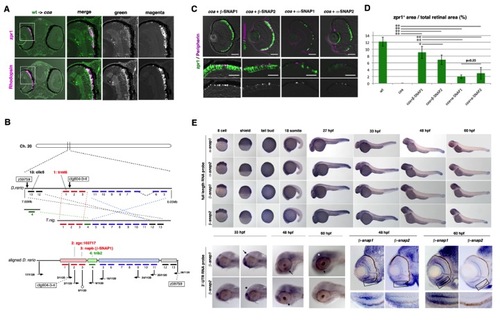
Cloning of the coa mutant gene, related to Figure 2
(A) Cell transplantation of wild-type donor cells into the coa mutant recipient retina. When wild-type cells labeled with biotin-dextran (green) were incorporated into the coa mutant retina, wild-type cells survived to express zpr1 (magenta, upper panels) and rhodopsin (magenta, lower panels) at 6 dpf. (B) The coa mutation is flanked by two polymorphic markers, ctg604-3-4 and z59759, on chromosome 20. These two markers are located close to the trmt6 and clic5 genes, respectively. On the Tetraodon nigroviridis genome, eleven genes are aligned between the trmt6 and clic5 genes (intermediate bar, T. nig.). On the other hand, 13 genes, including the trmt6 and clic5 genes, are shuffled in a different order on the zebrafish genome annotated in Ensemble Zv7 (version 50.7d) (top bar, D. rerio). We corrected the alignment of these genes on the zebrafish genome in accordance with the recombination rate of polymorphic markers (bottom bar, aligned D. rerio). As a result, the coa mutation mapped to the region between zgc:103717 and trib2, where β-snap1 is located. (C) Labeling of 84 hpf coa mutant retinas injected with DNA encoding heat-shock promoter-fused β-SNAP1, β-SNAP2, α-SNAP1 and α-SNAP2 with zpr1 antibody (green) and GFP-tagged peripherin or anti-rhodopsin antibody (magenta). GFP-tagged peripherin was normally localized in coa mutant photoreceptors expressing β-SNAP1. Similar to the case of β-SNAP1, photoreceptors were maintained and rhodopsin was normally localized in coa mutant photoreceptors expressing β-SNAP2. The maintenance of photoreceptors was partially rescued and some dotted signals for peripherin were observed in coa mutant photoreceptors expressing α-SNAP1 or α-SNAP2. Middle panels indicate higher magnification of the outer photoreceptor layer shown in upper panels. Lower panels show the magenta channel. GFP-tagged peripherin or rhodopsin (magenta) is transported to the apical region of photoreceptors in all cases. (D) The percentage of zpr1-positive region relative to the total retinal region. Green and black bars indicate the means and standard deviations, respectively. The percentage is 12% in wild type and nearly zero in the coa mutant. Photoreceptors are effectively rescued in coa mutant embryos expressing β-SNAP1 and β-SNAP2. Although photoreceptors are rescued in coa mutant embryos expressing α-SNAP1 and α-SNAP2, the percentages are lower than those in embryos expressing β-SNAP1 and β-SNAP2. Numbers of retinal sections used and p-values of t-tests are shown in Table S2. *p<0.05, **p<0.01. (E) Expression of α-snap1, α-snap2, β-snap1 and β-snap2 mRNA during development. Upper and lower panels indicate in situ hybridization using full-length and 3′-UTR RNA probes, respectively. All four mRNAs are expressed ubiquitously from eight cells to tail bud stage, and later restricted in the brain, especially the eye and tectum. At 33 hpf, β-snap1 mRNA is expressed in the telencephalon and pineal eye (arrow), whereas β-snap2 mRNA is not expressed in the pineal eye (asterisk). At 48 hpf, β-snap1 mRNA is expressed in the retina (arrows), whereas β-snap2 mRNA expression is not detected in the retina (asterisk). At 60 hpf, β-snap1 mRNA is expressed in the retina (arrow) and tectum (asterisk), whereas β-snap2 mRNA is very weak in both tissues. Sections of 48 and 60 hpf retinas labeled with β-snap1 and β-snap2 RNA probes show that β-snap1 mRNA is expressed in the retina, especially strong near the CMZ, but that β-snap2 mRNA expression is very weak in the retina. A higher magnification of the photoreceptor cell layer in the ventral retina is indicated. Scale bars: 50 μm (C, upper panels), 10 μm (C, middle panels)
|
