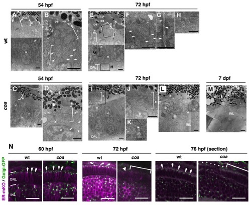
Amounts of OS and intracellular cisternae are reduced in coa mutant photoreceptors, related to Figure 1
(A, B) EM images of wild-type photoreceptors at 54 hpf. The OS starts to form. A mitochondria-rich region, which is called the ellipsoid (e), develops beneath the OS. Tubular cisternae (c) (B, arrows) are observed between the ellipsoid region (e) and nucleus (n). (C, D) EM images of coa mutant photoreceptors at 54 hpf. Ellipsoid region (e) and nuclei (n) are observed but the OS is absent or very small. Tubular cisternae are not well developed and small cisternae-like structures are observed near the nucleus (D, arrows). (E–H) EM images of wild-type photoreceptors at 72 hpf. The panels (F–H) indicate higher magnification. The OS becomes enlarged, and ellipsoid, tubular cisternae (F, arrows), nuclei and synapse ribbons (H) are observed. Tubular structures surrounding the nucleus, which possibly correspond to the ER, are observed (G, arrows). (I–L) EM images of coa mutant photoreceptors at 72 hpf. Panels (J, K) indicate higher magnification. Ellipsoid (e) and nucleus (n) are evident, but tubular cisternae structures are reduced between the ellipsoid and nucleus (J). Occasionally, mitochondria are associated with the nucleus (K, arrow). Some abnormal shaped nuclei and a phagosome-like structure containing mitochondria (arrowhead, L) are observed. (M) EM images of coa mutant photoreceptors at 7 dpf. The outer nuclear layer (ONL) is completely eliminated. The INL nuclei are associated with pigmented epithelium. (N) Confocal scanning of whole-mount (60 and 72 hpf) and sectioned (76 hpf) retinas of wild-type and coa mutant embryos injected with ER-mKO (magenta) and Golgi-GFP (green) mRNAs. ER-mKO and Golgi-GFP (arrows) are localized in the perinuclear region and the apical region of wild-type photoreceptors, respectively. In coa mutant photoreceptors, expression of both proteins is normal at 60 hpf, but Golgi-GFP signals are faint at 72 hpf (arrowhead) and subsequently disappear (bracket). A dotted line indicates degenerating photoreceptors in the coa mutant at 76 hpf. Abbreviations: c, tubular cisternae; e, ellipsoid; INL, inner plexiform layer; n, nucleus; OPL, outer plexiform layer; OS, outer segment. Scale bars: 1 μm (A–M), 10 μm (N).
|
