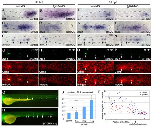
Knockdown of fgf10 delays initial neuromast formation and deposition. (A-H) Knockdown of fgf10 delays the initial restriction of the lef1 domain, the establishment of FGFR signaling and the formation of proto-neuromasts. lef1 (A,B), pea3 (C,D), and atoh1a (E,F) expression is shown in control (conMO) and fgf10 morphant embryos at 31 hpf. For statistical analysis, see supplementary material Fig. S8. (G,H) Epithelial rosette formation visualized by central accumulation (arrows) of ZO-1 (green) and Cldnb (red). (I-P) By 35 hpf, however, there is restriction of lef1 (I,J), recovery of pea3 (K,L) and establishment of multiple proto-neuromasts, as shown by focal atoh1a (M,N) and central accumulation of ZO-1 (green) and Cldnb (red) (O,P). (Q,R) Deposition of the first neuromast is delayed in fgf10 morphants and subsequent neuromasts are deposited at closer intervals. (S) Dose-dependent delay in L1 neuromast deposition. *P<0.05, ***P<0.001. Error bars indicate s.d. (T) Comparison of the relative size of the lef1 expression domain at different stages of migration in control (red) and fgf10 morphant (blue) embryos, as in Fig. 2E.
|
