
Fig. 1
- ID
- ZDB-FIG-120412-1
- Publication
- Nakao et al., 2012 - The role of mislocalized phototransduction in photoreceptor cell death of retinitis pigmentosa
- Other Figures
- All Figure Page
- Back to All Figure Page
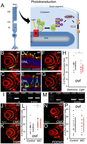
The pathway of photoreceptor cell death does not include PDE6β. (A) Schematic diagram of Phototransduction cascade. OS: outer segment, CC: connecting cilium, IS: inner segment, R: rhodopsin, T: transducin, PDE: phosphodiesterase (B and C) Eye ections of eyes from wt (B) and ovl (C) fish at 108 hpf. Rod photoreceptors are visualized with EGFP (Green). (Bar = 100 μm.) In ovl, the number of rod photoreceptors was decreased. (D and E) Transverse cryosections through wt (D) and ovl mutant (E) retinae at 4 dpf. F-actin is visualized with phalloidin (red), rod opsin with antibodies (green) and nuclei with Hoechst33342 (blue). Rhodopsin is mis-localized in ovl. Arrows indicate outer segments. (Bar = 10 μm.) ONL: outer nuclear layer, INL: inner nuclear layer (F and G)Ovl animals were reared in constant darkness (F) or in constant light (G) at 108 hpf. Light exposure reduces the survival of rod photoreceptor cells. (H) Graph of the number of rod photoreceptors in ovl fish at 108 hpf. (Bars mean SD, * means p<0.05.) (I) Trandsucin α expression analysis by RT-PCR of control morpholino- (lane1) and anti-Transducin α morpholino-treated (lane2). The expression of transducin is effectively suppressed by the morpholino at 108 hpf. CMO: control morpholino, TrMO: anti-Transducin α morpholino, (J and K) Sections of eyes treated by anti-transducin α morpholinos (K) and control MO (J) in ovl at 108 hpf. Anti-transducin a morpholinos rescued the rod photoreceptor cell death. (L) Rod photoreceptor numbers in anti-transducin α and control morpholino-treated ovl mutants at 108 hpf. (Bars mean SD, ** means p<0.01.) (M) PDE6β expression analysis by RT-PCR of control morpholino- (lane1) and anti-PDE6β (lane2) morpholino-treated mutants. The expression of PDE6β is effectively suppressed by the morpholino at 108 hpf. (N and O) Sections of eyes treated by anti-phosphodiesterase 6β morpholinos (O) and control MO (N) in ovl. (Bar = 100 μm.) There are no significant difference. (P) Rod photoreceptor survival in anti-phosphodiesterase 6β morpholino- (red dots) and control (black dots) morpholino-treated ovl mutants. Anti-PDE 6β morpholino has no significant effect on the number of rod photoreceptors. (Bars mean SD.) |
| Genes: | |
|---|---|
| Fish: | |
| Knockdown Reagents: | |
| Anatomical Term: | |
| Stage: | Day 4 |
| Fish: | |
|---|---|
| Condition: | |
| Knockdown Reagent: | |
| Observed In: | |
| Stage: | Day 4 |