FIGURE

Fig. S1

ID
ZDB-FIG-090617-52
Publication
Tucker et al., 2008 - The BMP signaling gradient patterns dorsoventral tissues in a temporally progressive manner along the anteroposterior axis
Other Figures
All Figure Page
Back to All Figure Page
Fig. S1

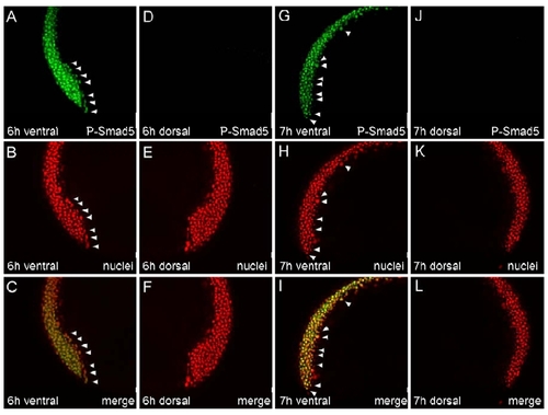
P-Smad5 Is Expressed in Ectodermal and Mesendodermal Cells on the Ventral Side of the Embryo during Gastrulation
(A, D, G, J) P-Smad5 expression. (B, E, H, K) Nuclear staining using TO-PRO-3 (Molecular Probes). (C, F, I, L) Overlay of above panels. P-Smad5 is expressed in nearly all cells on the ventral side of the embryo at all positions along the animal-vegetal axis at 6 hpf (A, B, C) and 7 hpf (G, H, I), but is not expressed in any cells on the dorsal side at the same time points (D, E, F, J, K, L). Arrowheads denote involuted mesendodermal cells expressing P-Smad5. All panels are 1 μM thick confocal slices, lateral views with animal pole to the top.

Expression Data

Expression Detail
Antibody Labeling
Phenotype Data

Phenotype Detail
Acknowledgments
This image is the copyrighted work of the attributed author or publisher, and ZFIN has permission only to display this image to its users. Additional permissions should be obtained from the applicable author or publisher of the image.

Reprinted from Developmental Cell, 14(1), Tucker, J.A., Mintzer, K.A., and Mullins, M.C., The BMP signaling gradient patterns dorsoventral tissues in a temporally progressive manner along the anteroposterior axis, 108-119, Copyright (2008) with permission from Elsevier. Full text @ Dev. Cell