
- Title
-
The BMP signaling gradient patterns dorsoventral tissues in a temporally progressive manner along the anteroposterior axis
- Authors
- Tucker, J.A., Mintzer, K.A., and Mullins, M.C.
- Source
- Full text @ Dev. Cell
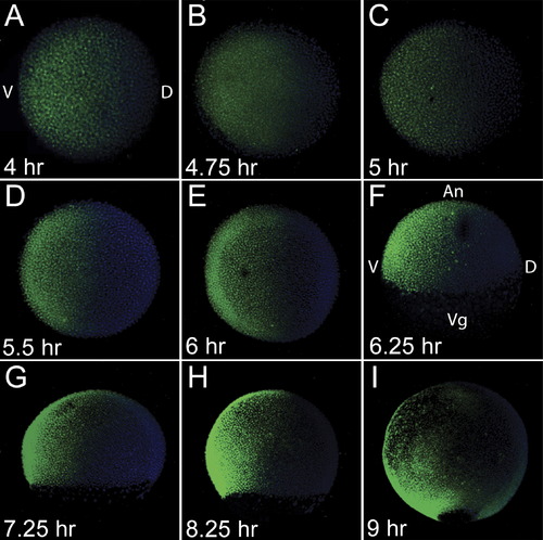
Formation of the BMP Activity Gradient Whole-mount P-Smad5 (green) antibody staining during blastula stages, at 4 hpf (A), 4.75 hpf (B), and 5 hpf (C) and during gastrula stages at 5.5 hpf (D), 6 hpf (E), 6.25 hpf (F), 7.25 hpf (G), 8.25 hpf (H), and 9 hpf (I). Nuclei were stained with DAPI (blue). (A)–(E) Animal view. (F)–(I) Lateral view. Dorsal to right in all. |
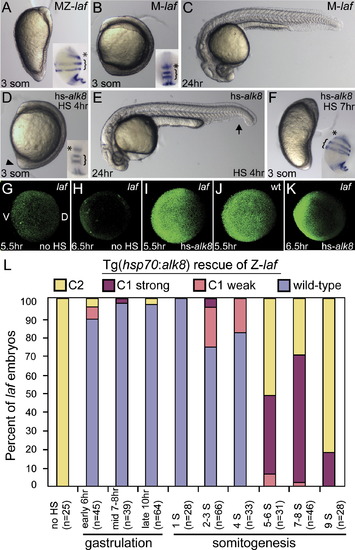
Temporal Series of Tg(hsp70:alk8) Induction to Rescue MZ-laf and Z-laf Live C5 dorsalized non-heat-shocked MZ-laf transgenic embryo at the three-somite (3 som) stage (A). Live M-laf embryos (laf/+ embryos from laf/laf mother) heat-shocked at 4 hpf display a wild-type (WT) phenotype at the three-somite stage (B) and at 24 hpf (C), consistent with a lack of alk8 overexpression phenotypic effects ([Bauer et al., 2001] and [Mintzer et al., 2001]). HS of MZ-laf; Tg(hsp70:alk8)/+ embryos at 4 hpf rescues them to C1 phenotype ([D], arrowhead marks protruding tail bud; [E], arrow marks partial loss of the ventral tail fin). HS at 7 hpf (F) fails to rescue MZ-laf transgenic embryos. MZ-laf mutants vary in the expansion of pax2.1 expression in the MHB (*), from greatly expanded ([A], inset) to circumferential ([F], inset). P-Smad5 in non-heat-shocked MZ-laf embryos at 5.5 hpf (G) and 6.5 hpf (H). After HS at 5 hpf, P-Smad5 at 5.5 hpf (I) and 6.5 hpf (K) in MZ-laf and WT (J). A higher gain was used to image all P-Smad5 embryos in this figure compared to Figure 1. (L) Graphic of extent of rescue of zygotic laf mutant transgenic embryos heat-shocked for 30 min at different time points. (G)–(K) are animal views, dorsal to right; insets in (B) and (D) are dorsal views, anterior to top; all others are lateral views, dorsal to right. Insets in (A), (B), (D), and (F) are pax2.1 (asterisk), krox20 (bracket), and myoD in situ hybridization at the five-somite stage. |

Tg(hsp70:chd) Rapidly Inhibits BMP Signaling, Generating a Range of Dorsalized Phenotypes Dependent on Induction Time (A) P-Smad5 western blot at blastula (3.0–5.25 hpf), onset of gastrulation (5.5 hpf) through midgastrulation (5.75–7.75 hpf) stages. (B) P-Smad5 during (20 and 40 min time points) and immediately after a 60 min HS at 4, 5, or 6 hpf. Actin is a loading control. (C–F) Nontransgenic WT (C) and transgenic siblings heat-shocked at 4 hpf (D) or 6 hpf (E) at the three-somite stage and at 1 day postfertilization (dpf) (F). (G and H) Dorsalization at 1 dpf of embryos heat-shocked at 7 hpf (G) and 10 hpf ([H], arrows indicate loss of ventral tail tissue). (I) Distribution of dorsalized phenotypes following HS at different time points. |

Tg(hsp70:chd)-Induced Dorsalization Is Apparent during Gastrulation Whole-mount in situ hybridization of nontransgenic heat-shocked WT siblings (A–D) compared to heat-shocked transgenic embryos (E–T). otx2 expression at 60%–65% epiboly in prospective anterior neural tissue in WT (A), or transgenic following HS at 4 hpf ([E], n = 9/9), 5 hpf ([I], n = 6/6), 5.5 hpf ([M], n = 19/19), or 6 hpf ([Q], n = 13/13). foxb1.2 expression in prospective neurectoderm in WT at 50% epiboly (B), or at 50% epiboly or shield stage immediately following HS at 4 hpf ([F], n = 3/3) or 5 hpf ([J], n = 9/9), respectively. foxb1.2 expression at 55%–60% epiboly following a 5.5 hpf HS ([N], n = 9/10), and at 65%–70% epiboly following a 6 hpf HS ([R], n = 15/15). AP-2 expression at 70% epiboly in ventral nonneural ectoderm in WT (C), and following HS at 4 hpf ([G], n = 13/13), 5 hpf ([K], n = 7/7), 5.5 hpf ([O], n = 7/7), or 6 hpf ([S], n = 8/8). dlx3 expression at 70% epiboly in ventral nonneural ectoderm in WT (D), and following HS at 4 hpf ([H], n = 12/12), 5 hpf ([L], n = 7/7), 5.5 hpf ([P], n = 8/10), or 6 hpf ([T], n = 12/16). gsc expression in dorsal midline mesoderm serves as a control for AP-2 and dlx-3, and is unaffected by HS. Arrowheads denote expression domains. (D), (H), (L), (P), and (T) are lateral views, dorsal to right; all others are animal views, dorsal to right. |
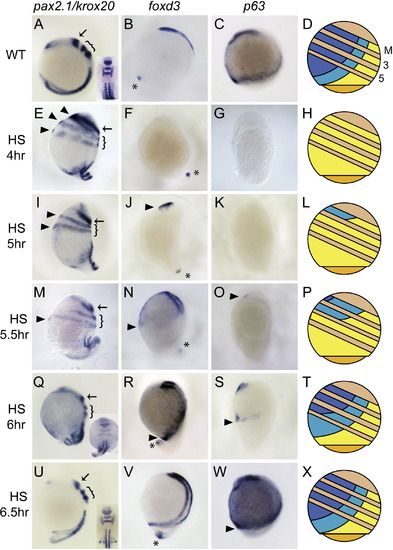
Progressive BMP Inhibition Series by Tg(hsp70:chd) Reveals Progressive DV Ectodermal Patterning along the AP Axis Expression of pax2.1 in the MHB (arrow), krox20 in R3 and R5 (bracket), and myoD in somitic mesoderm at the six-somite stage in WT (A), and following HS at 4 hpf ([E], n = 17/19), 5 hpf ([I], n = 15/21), 5.5 hpf ([M], n = 13/18), 6 hpf ([Q], n = 21/21), or 6.5 hpf ([U], n = 11/11). Arrowheads denote circumferential neural tissues. foxd3 expression in CNC at the six-somite stage in WT (B), and following HS at 4 hpf ([F], n = 12/13), 5 hpf ([J], n = 13/21), 5.5 hpf ([N], n = 15/19), 6 hpf ([R], n = 11/15), or 6.5 hpf (V). Asterisks denote unaffected tail bud expression. p63 expression in prospective epidermis at the two-somite stage in WT (C), and following HS at 4 hpf ([G], n = 8/8), 5 hpf ([K], n = 16/16), 5.5 hpf ([O], n = 8/17), 6 hpf ([S], n = 16/24), or 6.5 hpf ([W], n = 5/5). (D, H, L, P, T, and X) Representations summarize the relative domains of the three tissue types. Yellow, neurectoderm; turquoise, neural crest; blue, epidermis. M indicates MHB boundary; 3 and 5 are R3 and R5. Tan stripes are the regions between the MHB, R3, and R5 AP positions across the DV axis. All are lateral views, dorsal to right, anterior to top, except for insets in (A), (Q), and (U), which are dorsal views, anterior to top. |
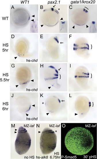
Progressive BMP Inhibition Series Reveals Progressive DV Mesodermal Patterning along the AP Axis, and Rescue of Posterior, but Not Anterior, in MZ-laf Embryos (A–L) WT1 expression in the pronephros in heat-shocked nontransgenic WT siblings (A), and following HS at 5 hpf ([D], n = 16/19), 5.5 hpf ([G], n = 7/12), or 6 hpf ([J], n = 10/12). pax2.1 expression in pronephros (arrowheads), MHB (arrow), and otic placode (asterisk) in WT (B), and following HS at 5 hpf ([E], n = 16/16), 5.5 hpf ([H], n = 14/24), or 6 hpf ([K], n = 10/13). krox20 expression was also examined in ([H], bracket). Expression of gata1 in blood progenitors (arrowheads) and krox20 in hindbrain (brackets) in WT (C), and following HS at 5 hpf ([F], n = 5/5), 5.5 hpf ([I], n = 6/6), or 6 hpf ([L], n = 5/7). (M and N) krox20 expression in R3 and R5 (arrowheads), and myoD in somitic mesoderm in MZ-laf mutants overexpressing bmp2b in the absence of HS (M) or following a 30 min heat-shock induction at 6.75 hpf (N) of Tg(hsp70:alk8) causes partial expansion of R5 (asterisk) but complete expansion of R3 (arrowhead), and reinitiation of P-Smad5 by 7.25 hpf (O). (A)–(N) are lateral views, dorsal to right, at the five-somite stage. (O) is animal pole view, dorsal to right. |
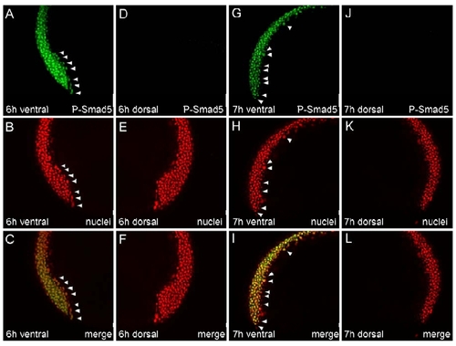
P-Smad5 Is Expressed in Ectodermal and Mesendodermal Cells on the Ventral Side of the Embryo during Gastrulation |
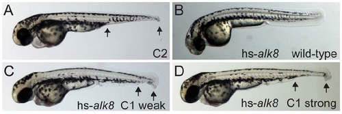
Tg(hsp70:alk8) Rescues Zygotic laf Mutants during Early Somitogenesis |
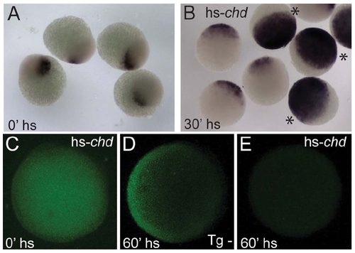
BMP Signaling through Smad5 Is Rapidly Inhibited by Tg(hsp70:chd) Induction |
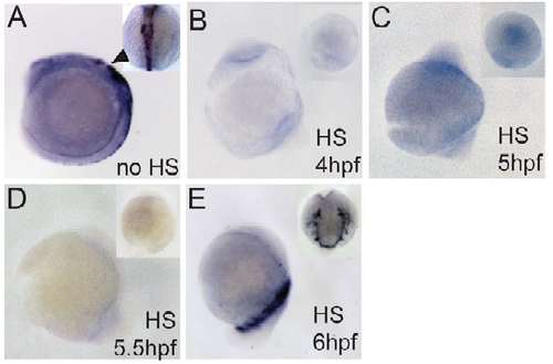
crestin Expression in Trunk Neural Crest Following Tg(hsp70:chd) Induction and BMP Inhibition |
Reprinted from Developmental Cell, 14(1), Tucker, J.A., Mintzer, K.A., and Mullins, M.C., The BMP signaling gradient patterns dorsoventral tissues in a temporally progressive manner along the anteroposterior axis, 108-119, Copyright (2008) with permission from Elsevier. Full text @ Dev. Cell