
Fig. 2
- ID
- ZDB-FIG-090617-47
- Publication
- Tucker et al., 2008 - The BMP signaling gradient patterns dorsoventral tissues in a temporally progressive manner along the anteroposterior axis
- Other Figures
- All Figure Page
- Back to All Figure Page
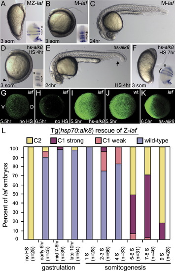
Temporal Series of Tg(hsp70:alk8) Induction to Rescue MZ-laf and Z-laf Live C5 dorsalized non-heat-shocked MZ-laf transgenic embryo at the three-somite (3 som) stage (A). Live M-laf embryos (laf/+ embryos from laf/laf mother) heat-shocked at 4 hpf display a wild-type (WT) phenotype at the three-somite stage (B) and at 24 hpf (C), consistent with a lack of alk8 overexpression phenotypic effects ([Bauer et al., 2001] and [Mintzer et al., 2001]). HS of MZ-laf; Tg(hsp70:alk8)/+ embryos at 4 hpf rescues them to C1 phenotype ([D], arrowhead marks protruding tail bud; [E], arrow marks partial loss of the ventral tail fin). HS at 7 hpf (F) fails to rescue MZ-laf transgenic embryos. MZ-laf mutants vary in the expansion of pax2.1 expression in the MHB (*), from greatly expanded ([A], inset) to circumferential ([F], inset). P-Smad5 in non-heat-shocked MZ-laf embryos at 5.5 hpf (G) and 6.5 hpf (H). After HS at 5 hpf, P-Smad5 at 5.5 hpf (I) and 6.5 hpf (K) in MZ-laf and WT (J). A higher gain was used to image all P-Smad5 embryos in this figure compared to Figure 1. (L) Graphic of extent of rescue of zygotic laf mutant transgenic embryos heat-shocked for 30 min at different time points. (G)–(K) are animal views, dorsal to right; insets in (B) and (D) are dorsal views, anterior to top; all others are lateral views, dorsal to right. Insets in (A), (B), (D), and (F) are pax2.1 (asterisk), krox20 (bracket), and myoD in situ hybridization at the five-somite stage. |
Reprinted from Developmental Cell, 14(1), Tucker, J.A., Mintzer, K.A., and Mullins, M.C., The BMP signaling gradient patterns dorsoventral tissues in a temporally progressive manner along the anteroposterior axis, 108-119, Copyright (2008) with permission from Elsevier. Full text @ Dev. Cell