
Fig. 5
- ID
- ZDB-FIG-090617-50
- Publication
- Tucker et al., 2008 - The BMP signaling gradient patterns dorsoventral tissues in a temporally progressive manner along the anteroposterior axis
- Other Figures
- All Figure Page
- Back to All Figure Page
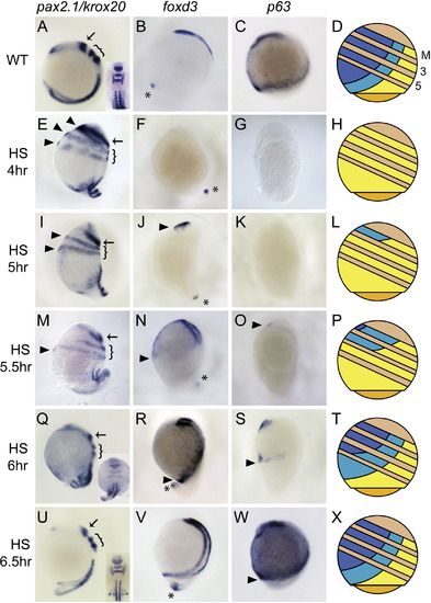
Progressive BMP Inhibition Series by Tg(hsp70:chd) Reveals Progressive DV Ectodermal Patterning along the AP Axis Expression of pax2.1 in the MHB (arrow), krox20 in R3 and R5 (bracket), and myoD in somitic mesoderm at the six-somite stage in WT (A), and following HS at 4 hpf ([E], n = 17/19), 5 hpf ([I], n = 15/21), 5.5 hpf ([M], n = 13/18), 6 hpf ([Q], n = 21/21), or 6.5 hpf ([U], n = 11/11). Arrowheads denote circumferential neural tissues. foxd3 expression in CNC at the six-somite stage in WT (B), and following HS at 4 hpf ([F], n = 12/13), 5 hpf ([J], n = 13/21), 5.5 hpf ([N], n = 15/19), 6 hpf ([R], n = 11/15), or 6.5 hpf (V). Asterisks denote unaffected tail bud expression. p63 expression in prospective epidermis at the two-somite stage in WT (C), and following HS at 4 hpf ([G], n = 8/8), 5 hpf ([K], n = 16/16), 5.5 hpf ([O], n = 8/17), 6 hpf ([S], n = 16/24), or 6.5 hpf ([W], n = 5/5). (D, H, L, P, T, and X) Representations summarize the relative domains of the three tissue types. Yellow, neurectoderm; turquoise, neural crest; blue, epidermis. M indicates MHB boundary; 3 and 5 are R3 and R5. Tan stripes are the regions between the MHB, R3, and R5 AP positions across the DV axis. All are lateral views, dorsal to right, anterior to top, except for insets in (A), (Q), and (U), which are dorsal views, anterior to top. |
Reprinted from Developmental Cell, 14(1), Tucker, J.A., Mintzer, K.A., and Mullins, M.C., The BMP signaling gradient patterns dorsoventral tissues in a temporally progressive manner along the anteroposterior axis, 108-119, Copyright (2008) with permission from Elsevier. Full text @ Dev. Cell