
Fig. S7
- ID
- ZDB-FIG-090501-71
- Publication
- de Pater et al., 2009 - Distinct phases of cardiomyocyte differentiation regulate growth of the zebrafish heart
- Other Figures
- All Figure Page
- Back to All Figure Page
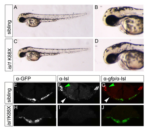
isl1K88X mutant embryos are deficient for Isl protein. (A-D) Live images of 48-hpf isl1K88X sibling and mutant embryos showing no apparent morphological phenotype. (E-J) Single z-scans of confocal images of 23-somite stage isl1K88X Tg(cmlc2:eGFP) embryos after immunofluorescence staining with α-eGFP (E,H) and α-Isl (F,I) antibodies. (E-G) isl1K88X sibling embryo; (H-J) isl1K88X mutant embryo. Arrowheads in the overlay of α-eGFP (green) with α-Isl (red) indicate the eGFPposIslpos cells in the cardiac disk and trigeminal sensory ganglia. In the control (E-G), Isl is expressed in cardiac cells located at lateral positions in the cardiac disk (white arrowhead) and in the trigeminal sensory ganglion (green arrowhead), described in Fig. 3A-C. Note that the nuclear Isl signal in the cardiac disk in H-J is absent. Scale bars: 80 μm. |