
FIGURE
Fig. S1
- ID
- ZDB-FIG-090501-66
- Publication
- de Pater et al., 2009 - Distinct phases of cardiomyocyte differentiation regulate growth of the zebrafish heart
- Other Figures
- All Figure Page
- Back to All Figure Page
Fig. S1
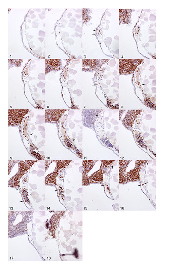
BrdU pulse labeling of cardiac cells. Sagittal sections of a single embryo after pulse labeling with BrdU from 24-48 hpf. The sections were immunohistochemically stained with an α-BrdU antibody (brown) and counter-stained with Hematoxylin and Eosin (blue). Arrows indicate BrdU-positive myocardial cells. Note the high degree of labeling within the embryonic blood cells and endocardial cells. Anterior is to the left, dorsal to the top. |
Expression Data
Expression Detail
Antibody Labeling
Phenotype Data
Phenotype Detail
Acknowledgments
This image is the copyrighted work of the attributed author or publisher, and
ZFIN has permission only to display this image to its users.
Additional permissions should be obtained from the applicable author or publisher of the image.
Full text @ Development