
Fig. S5
- ID
- ZDB-FIG-090501-70
- Publication
- de Pater et al., 2009 - Distinct phases of cardiomyocyte differentiation regulate growth of the zebrafish heart
- Other Figures
- All Figure Page
- Back to All Figure Page
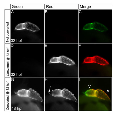
Photoconversion of Kaede efficiently marks differentiated cardiomyocytes. (A-I) Lateral views of Tg(cmlc2:kaede) embryos, ventricle to the left, demonstrating effectiveness of photoconversion technique. (A-C) Prior to photoconversion, the 32-hpf heart exhibits green Kaede fluorescence (A) but not red Kaede fluorescence (B), resulting in an entirely green heart (C). (D-F) Immediately after exposure to UV light, all of the green Kaede (D) has been converted into red Kaede (E), resulting in an entirely red heart (F). (G-I) Sixteen hours after photoconversion, red Kaede (H) persists in the cells that were expressing the transgene. These cells also generate new green Kaede (G), as do newly differentiating cells that initiated transgene expression after 32 hpf. As a result, cardiomyocytes at the arterial pole fluoresce green, but not red, at 48 hpf (arrow in H). V, ventricle; A, atrium. |