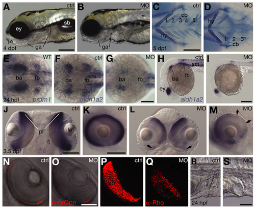
Additional organogenesis defects resulting from downregulation of Prdm1 activity in branchial arches, eyes and cloaca. (A,B,H,I,K,M,R,S) Lateral view, anterior towards the left. (C,D) Ventral view, anterior towards the left. (E-G) Dorsal view, anterior towards the left. (J) Dorsal view, anterior towards the top. (L) Ventral view, anterior towards the top. (N-Q) Ventral view, anterior towards top right, left eye. (J,K and L,M) Different views of the same embryos. (A,C,E,F,H,J,K,N,P,R) Untreated controls (ctrl). (B,D,G,I,L,M,O,Q,S) Embryos microinjected with 1 ng of MOprdm1 oligonucleotides (MO). (A,B) Morphants exhibit jaw (jw) and gill arch defects (ga), and lack inflated swim bladders (sb); eye (ey). (C,D) Development of pharyngeal skeleton, visualized by Alcian Blue cartilage staining in 5 dpf morphants (Piotrowski et al. 1996); hyoid (hy) is abnormal, ceratobranchials 1-3 (cb) are shortened and ceratobranchials 4-5 are missing (class III, 24%; class II morphants lack cb 3-5 and class I lack cb1/2-5; 63% and 13%, respectively, n=215). (E,F) prdm1 and aldh1a2 share expression domains in the branchial arch (ba) and pectoral fin bud (fb) primordia (Grandel et al., 2002). (F-I) Morphant embryos exhibit reduced aldh1a2 expression in branchial arch and pectoral fin primordia; and eyes (ey) and somites (sm). (J-M) Eye size and rho expression in photoreceptor cell layer (pl) (Vihtelic et al., 1999) is reduced in morphants; lens (le) and retina lamination (rt) appear normal, whereas rho expression is generally reduced and missing regionally (arrows). (N,O) Reduced number of green-sensitive Opsin-expressing cells in PCL of morphants, detected by a-grOpn antibody (Vihtelic et al., 1999); single focal level obtained by confocal microscopy. (P,Q) Strongly reduced number of Rhodopsin-expressing cells in PCL of morphants, detected by a-Rho antibody (Zpr3/Fret11, Vihtelic et al., 1999); images obtained by 3D reconstruction. Morphant eye dimensions are smaller. Cy3-conjugated secondary antibodies by Jackson ImmunoResearch were used. (R,S) Cloaca (cl) morphology is abnormal in morphants. Scale bar: 200 µm in A,C,I; 100 µm in G,J,K; 50 µm in O,U.
|
