- Title
-
Induction of the mesendoderm in the zebrafish germ ring by yolk cell-derived TGF-ß; family signals and discrimination of mesoderm and endoderm by FGF
- Authors
- Rodaway, A., Takeda, H., Koshida, S., Broadbent, J., Price, B., Smith, J.C., Patient, R., and Holder, N.
- Source
- Full text @ Development
|
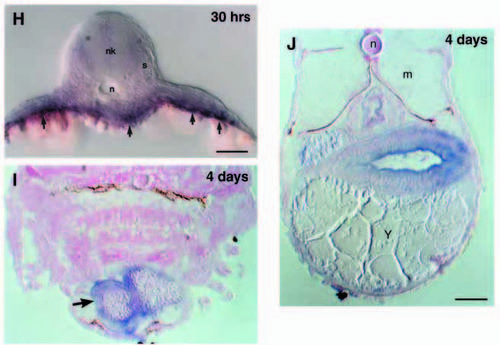
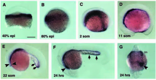
Expression of zebrafish gata5 detected by in situ hybridisation. Stages according to Kimmel et al. (1995). In A and B the animal pole is up and in C-F anterior is to the left and dorsal is up. (A) 40% epiboly. gata5 expression in the germ ring. (B) 80% epiboly. gata5-expressing cells have involuted and dispersed towards the animal pole. (C) 2 somites. gata5-expressing cells are in the lateral mesoderm anteriorly, and in presumptive endoderm cells converging towards the midline. (D) 11 somites. The anterior extent of the expressing cells is beneath the midbrain. (E) 22 somites. The heart is expressing gata5 (arrowheads). Caudally, expressing cells are present around the yolk cell extension (arrows). (F) 24 hours. The arrowhead marks the heart and the arrows expression around the yolk cell extension. (G) 24 hour, frontal view. gata5 expression in the heart on the left hand side of the embryo (arrowhead). More caudally, expression is across the midline in a thin layer of presumptive endoderm, and in the anterior lateral mesoderm (lm). (H) 30 hours (thick section). Cells expressing gata5 are close to the yolk cell (arrows). neural keel (nk); notochord (n); somites (s). At 4 days (10 mm sections) gata5 expression is maintained in the heart (arrow in I) and the gut (J). Labels in J are n, notochord; m, muscle; y, yolk. Scale bars, 250 μm (A-G); 50 μm (H); 25 μm (I,J). EXPRESSION / LABELING:
|
|
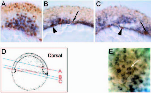
gata5 is expressed in endodermal cells in the gastrula embryo. (A-C) Cryostat sections of a 60% epiboly embryo double stained for gata5 message (in situ hybridisation; blue cytoplasmic staining) and fkd2 protein (antibody; brown nuclear staining). (A) Dorsal view. Coexpression of gata5 and fkd2 in the mesendoderm of the prechordal plate. (B) Lateral view. Coexpression of gata5 and fkd2 in endodermal precursors adjacent to the yolk cell, in the innermost layer of the hypoblast (arrow). fkd2 also expressed in the YSL (arrowhead) which weakly expresses gata5. (C) Ventral view. fkd2 is expressed predominantly in the YSL (arrowhead) with only the occasional hypoblast cell, coexpressing gata5 (arrow). gata5 is expressed in several layers of hypoblast cells without coexpression of fkd2. These may include precursors of the anterior lateral mesoderm. (D) Diagram showing the plane of sections (blue lines) and position in sections (red lines) in parts A-C. (E) High power lateral view of 80% epiboly embryo whole-mount focused immediately above the yolk cell. gata5 is expressed in cells (arrowed) having the characteristic position (adjacent to the yolk cell) and shape (large, flat cells) of endodermal precursors (Warga and Nusslein-Volhard, 1999). EXPRESSION / LABELING:
|
|
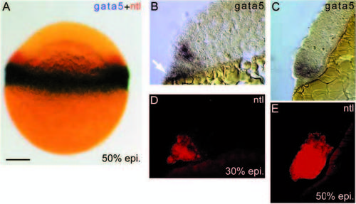
gata5 and ntl coexpress in the blastoderm margin. (A) Twocolour whole-mount in situ of a 50% epiboly embryo showing gata5 (blue) expression in the margin of the blastoderm, and wider expression of ntl (red). Scale bar,150 μm. (B-E) sequential in situ detection on the same sections of gata5 (blue; B and C) and ntl (red fluorescence; D and E), at 30% epiboly (B,D) and 50% epiboly (C,E). (B) The initial expression of gata5 in the blastoderm extends 2-3 cell diameters from the blastoderm margin. Expression is also detected in the YSL (white arrow). (C) At 50% epiboly, gata5 expresses for approximately the same distance from the blastoderm margin, which now represents 3-4 cell diameters. (D) Initially ntl expresses in the same blastomeres as gata5, but not in the YSL. (E) 50% epiboly. ntl expression has spread to encompass blastomeres within 8-10 cell diameters of the margin. Thus, by the beginning of gastrulation, gata5-expressing cells are a marginal subset of those expressing ntl. EXPRESSION / LABELING:
|
|
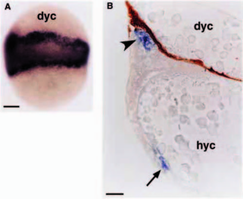
Induction of gata5 by a signal from the yolk cell. (A) Embryo at 60% epiboly with a grafted second yolk cell. gata5 is expressed in cells in association with both the host and the donor yolk cell (dyc). (B) A section through such a grafted embryo showing the additional layer of gata5-expressing cells (arrowhead). Normal gata5 expression in the host embryo is arrowed. The dark material in the donor yolk cell is dye injected prior to grafting to distinguish it from the host yolk cell (hyc). Scale bars, 150 μm (A); 40 μm (B). |
|
Patterned response of gata5 and ntl expression to activin; dependence of both genes on TGF-β signalling. (A-D) 50% epiboly embryo coinjected at 16-32 cell stage with activin (10 mg/ml) and nls-β-gal (100 mg/ml) RNA. β-gal detected with X-Gal (turquoise nuclear staining). The embryo was photographed after staining for ntl (red) and again after detection of gata5 (blue; B,D). (A,B) Low magnification, animal pole views of the whole embryo. (C,D) Higher magnification views of the patch of ectopic expression. (C) ntl expression overlaps with the injected cells and extends 4-8 cell diameters beyond the cells injected. (D) gata5 is induced in injected cells and up to 2-3 cell diameters beyond them. A ring of cells expressing ntl alone extends for about 4 cell diameters beyond those coexpressing ntl and gata5, mimicking the normal pattern of expression of these genes. (E-G) Blastoderm margin, 50% epiboly embryos coinjected at 2-cell stage with 1 mg/ml dnXAR and 100 ng/ml nls-β-gal RNA. (E) Stained for ntl expression (red); (F) stained for gata5 expression (blue) after staining for β-gal activity and (G) gata5 stain without staining for β-gal to show more clearly the gap in gata5 expression. Expression of both genes is eliminated by inhibiting TGF-β signalling. |
|
Induction of gata5 and ntl in embryos injected with RNA encoding constitutively active activin receptor, Alk4* (A-D) or BMP2/7 receptor, Alk2* (E), animal pole views, 50% epiboly. ntl, (red); gata5, (blue); b-gal, (turquoise). (A) 1 mg/ml Alk-4*. There is little alteration to the expression of ntl and gata5. (B) 10 μg/ml Alk4*. The expression of ntl and gata5 is ectopic in the location of cells expressing b-gal. gata5 ectopic expression occurs within the wider area of ntl expression. (C) 100 μg/ml of Alk4*. ntl expression is clearly in regions of the blastula not expressing β-gal whilst gata5 expression remains in the β-gal-expressing cells. This is shown in higher magnification in D. Black arrowhead indicates ntl-expressing cells without nuclear β-gal. gata5 expression is seen as blue cytoplasm in cells with turquoise nuclei (white arrows). (E) 100 μg/ml Alk2*. Normal expression of ntl and gata5. |
|
Ntl but not gata5 is positively regulated by FGF. A,C,E are 50% epiboly embryos hybridised with a probe to ntl. (A) Uninjected (uninj) embryo. (C) Injection of eFGF RNA causes widespread expression of ntl. (E) Injection of RNA encoding dominant negative FGF receptor, XFD. Where the β-gal tracer overlaps the germ ring, ntl expression is abolished in all but a single tier of cells closest to the blastoderm margin. B,D,F are embryos hybridised to gata5 probe. (B) uninjected embryo, (D) injected with eFGF, (F) injected with XFD. Neither eFGF nor XFD affect gata5 expression. Scale bar, 150 μm. EXPRESSION / LABELING:
|
|
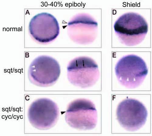
gata5 expression phenotypes obtained from a cross of fish doubly heterozygotic for squint (sqt cz35) and cyclops (cycm294). Expression was assayed at 30-40% epiboly (A-C, left embryos oblique animal pole view, right embryos lateral view) and shield stage (D-F, animal-lateral view). A and D show the normal expression pattern seen in most embryos. (A) At 30-40% epiboly, expression is in the margin of the blastoderm (open arrowhead) and the underlying YSL (black arrowhead). At shield stage (D) gata5 is expressed in the newly involuted hypoblast. One abnormal phenotype (B and E) was the same as that found in embryos from crosses of sqt single heterozygotes. At 30-40% epiboly (B), expression of gata5 in the blastoderm margin was generally weaker (black arrows) and in some embryos expression was absent from part of the blastoderm margin (white arrowheads). At shield stage (E), expression was absent from typically one quarter of the hypoblast (white arrowheads), and reduced in the remainder. In the second abnormal phenotype, representing embryos homozygous for both sqtcz35 and cycm294, gata5 was completely absent from blastomeres at both stages (C and F). Some remaining expression could be seen in the YSL (C, black arrowhead). EXPRESSION / LABELING:
|

