|
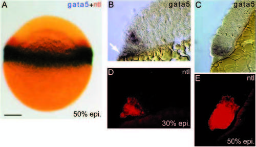
gata5 and ntl coexpress in the blastoderm margin. (A) Twocolour whole-mount in situ of a 50% epiboly embryo showing gata5 (blue) expression in the margin of the blastoderm, and wider expression of ntl (red). Scale bar,150 μm. (B-E) sequential in situ detection on the same sections of gata5 (blue; B and C) and ntl (red fluorescence; D and E), at 30% epiboly (B,D) and 50% epiboly (C,E). (B) The initial expression of gata5 in the blastoderm extends 2-3 cell diameters from the blastoderm margin. Expression is also detected in the YSL (white arrow). (C) At 50% epiboly, gata5 expresses for approximately the same distance from the blastoderm margin, which now represents 3-4 cell diameters. (D) Initially ntl expresses in the same blastomeres as gata5, but not in the YSL. (E) 50% epiboly. ntl expression has spread to encompass blastomeres within 8-10 cell diameters of the margin. Thus, by the beginning of gastrulation, gata5-expressing cells are a marginal subset of those expressing ntl.
|