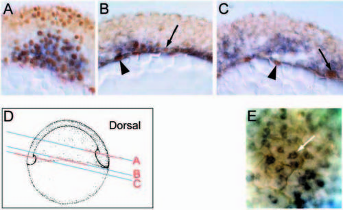
gata5 is expressed in endodermal cells in the gastrula embryo. (A-C) Cryostat sections of a 60% epiboly embryo double stained for gata5 message (in situ hybridisation; blue cytoplasmic staining) and fkd2 protein (antibody; brown nuclear staining). (A) Dorsal view. Coexpression of gata5 and fkd2 in the mesendoderm of the prechordal plate. (B) Lateral view. Coexpression of gata5 and fkd2 in endodermal precursors adjacent to the yolk cell, in the innermost layer of the hypoblast (arrow). fkd2 also expressed in the YSL (arrowhead) which weakly expresses gata5. (C) Ventral view. fkd2 is expressed predominantly in the YSL (arrowhead) with only the occasional hypoblast cell, coexpressing gata5 (arrow). gata5 is expressed in several layers of hypoblast cells without coexpression of fkd2. These may include precursors of the anterior lateral mesoderm. (D) Diagram showing the plane of sections (blue lines) and position in sections (red lines) in parts A-C. (E) High power lateral view of 80% epiboly embryo whole-mount focused immediately above the yolk cell. gata5 is expressed in cells (arrowed) having the characteristic position (adjacent to the yolk cell) and shape (large, flat cells) of endodermal precursors (Warga and Nusslein-Volhard, 1999).
|
