
- Title
-
Five Trk receptors in the zebrafish
- Authors
- Martin, S.C., Marazzi, G., Sandell, J.H., and Heinrich, G.
- Source
- Full text @ Dev. Biol.
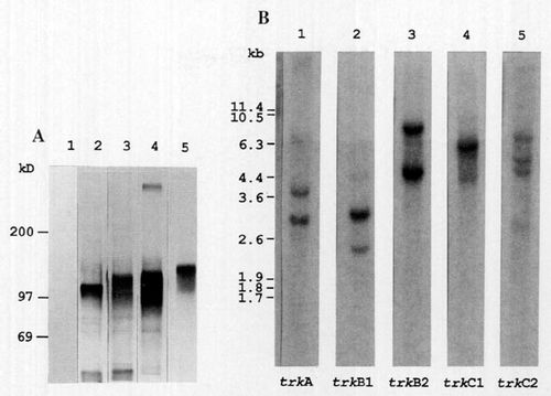
Immunoprecipitation of zebrafish Trk RTKs and Southern blot analysis of zebrafish trk genes. (A) Zebrafish Trk proteins were expressed in COS-1 cells and immunoprecipitated with anti-Trk serum. The immunoprecipitates were subjected to SDS-PAGE and exposed to X-ray film. Lane 1, mock transfected; lane 2, rat trkA; lane 3, trkB1; lane 4, trkB2; lane 5, trkC1. The positions of protein standards of the indicated molecular weights in kilodaltons are shown on the left. (B) Zebrafish genomic DNA was digested with BglII and examined by Southern blot hybridization. The probes were the products of PCR III. The probe region contains a BglII site in trkB1 and trkB2. Hybridizations were carried out in 2x SSC at 68°C. The identity of each probe is indicated below the lanes. The sizes in kilobases of bacteriophage λ Cla1 fragments are shown on the left. |
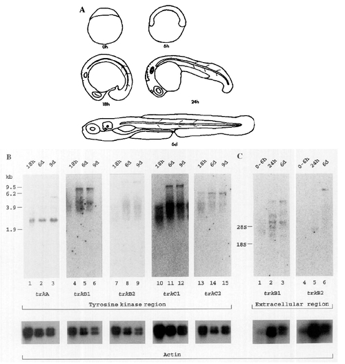
Northern blot analysis of zebrafish trk expression. (A) Diagrammatic outline of zebrafish developmental stages at the indicated stages in hours and days after fertilization, (B and C) Aliquots of 10 μg (B) or 2 μg (C) of poly(A)+ RNA from the time after fertilization is indicated above each lane. Hybridizations were carried out at 68°C in 2x SSC. The identity of the trk probe used is indicated below each panel. Different size markers were used for the blots in B and C. RNA markers for B are in kilobases. The positions of 28S and 18S rRNAs are marked for C. The lower panel is the result of hybridizations of each filter to chicken β-actin. The data were collected with a Fuji BAS 2000 Bioimager, with the exception of the actin signals that were recorded on X-ray film. |
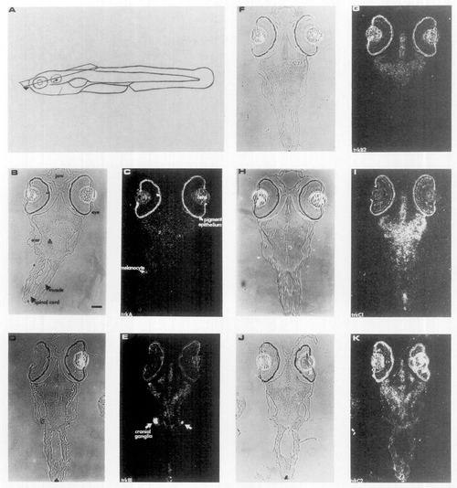
Zebrafish trk genes are expressed in the central and peripheral nervous system. Five RNA probes labeled with [35S]UTP specific for the full-length transcripts of the trk genes were used to examine expression 6 days after fertilization by in situ hybridization. (A) Schematic indicating the most dorsal plane of the sections of the zebrafish embryo. The position of the subsequent sections is indicated by the arrowhead. (C) trkA, (E) trkB1, (G) trkB2, (I) trkC1, and (J) trkC2 are dark-field photomicrographs. B, D, F, H, and J are phase photomicrographs of C, E, G, I, and K, respectively. The m and arrowhead in B marks the cell body of the Mauthner cell. The retinal pigmented epithelium melanocytes and iridiophores contain a chromophore which causes the formation of silver grains in the photographic emulsion and is not hybridization signal. Such a melanocyte (or iridiophore) is labeled with an arrow in C to illustrate this phenomenon. The lens appears white, but this is not hybridization signal, as it appeared the same without hybridization. The scale bar is 50 μm. EXPRESSION / LABELING:
|
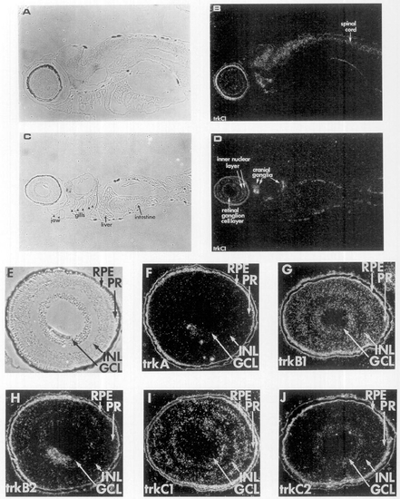
Zebrafish trk expression in the spinal cord, cranial ganglia, and retina. RNA probes labeled with [35S]UTP specific for trk full-length transcripts were used to examine expression 6 days after fertilization, in sagittal sections for trkC1 (A-D) and the retina for the five trk probes (E-J) using in situ hybridization. (B and D) trkC1 dark-field photomicrographs. (A and C) phase photomicrographs of B and D, respectively. (F) trkA, (G) trkB1, (H) trkB2, (I) trkC1, and (J) trkC2 are dark-field photomicrographs. (E) Phase photomicrograph of F (trkA). The positions of retinal pigment epithelium (RPE), photoreceptors (PR), inner nuclear layer (INL), and ganglion cell layer (GCL) are indicated. The lens next to the GCL in F and H appears white but does not correspond to labeling. The scale bar is 50 μm. EXPRESSION / LABELING:
|
Reprinted from Developmental Biology, 169, Martin, S.C., Marazzi, G., Sandell, J.H., and Heinrich, G., Five Trk receptors in the zebrafish, 745-758, Copyright (1995) with permission from Elsevier. Full text @ Dev. Biol.