
Fig. 4
- ID
- ZDB-FIG-090515-35
- Publication
- Martin et al., 1995 - Five Trk receptors in the zebrafish
- Other Figures
- All Figure Page
- Back to All Figure Page
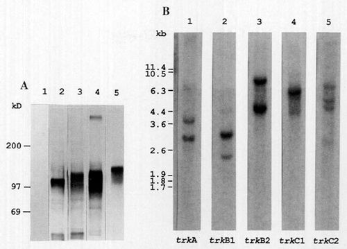
Immunoprecipitation of zebrafish Trk RTKs and Southern blot analysis of zebrafish trk genes. (A) Zebrafish Trk proteins were expressed in COS-1 cells and immunoprecipitated with anti-Trk serum. The immunoprecipitates were subjected to SDS-PAGE and exposed to X-ray film. Lane 1, mock transfected; lane 2, rat trkA; lane 3, trkB1; lane 4, trkB2; lane 5, trkC1. The positions of protein standards of the indicated molecular weights in kilodaltons are shown on the left. (B) Zebrafish genomic DNA was digested with BglII and examined by Southern blot hybridization. The probes were the products of PCR III. The probe region contains a BglII site in trkB1 and trkB2. Hybridizations were carried out in 2x SSC at 68°C. The identity of each probe is indicated below the lanes. The sizes in kilobases of bacteriophage λ Cla1 fragments are shown on the left. |
Reprinted from Developmental Biology, 169, Martin, S.C., Marazzi, G., Sandell, J.H., and Heinrich, G., Five Trk receptors in the zebrafish, 745-758, Copyright (1995) with permission from Elsevier. Full text @ Dev. Biol.