
Fig. 7
|
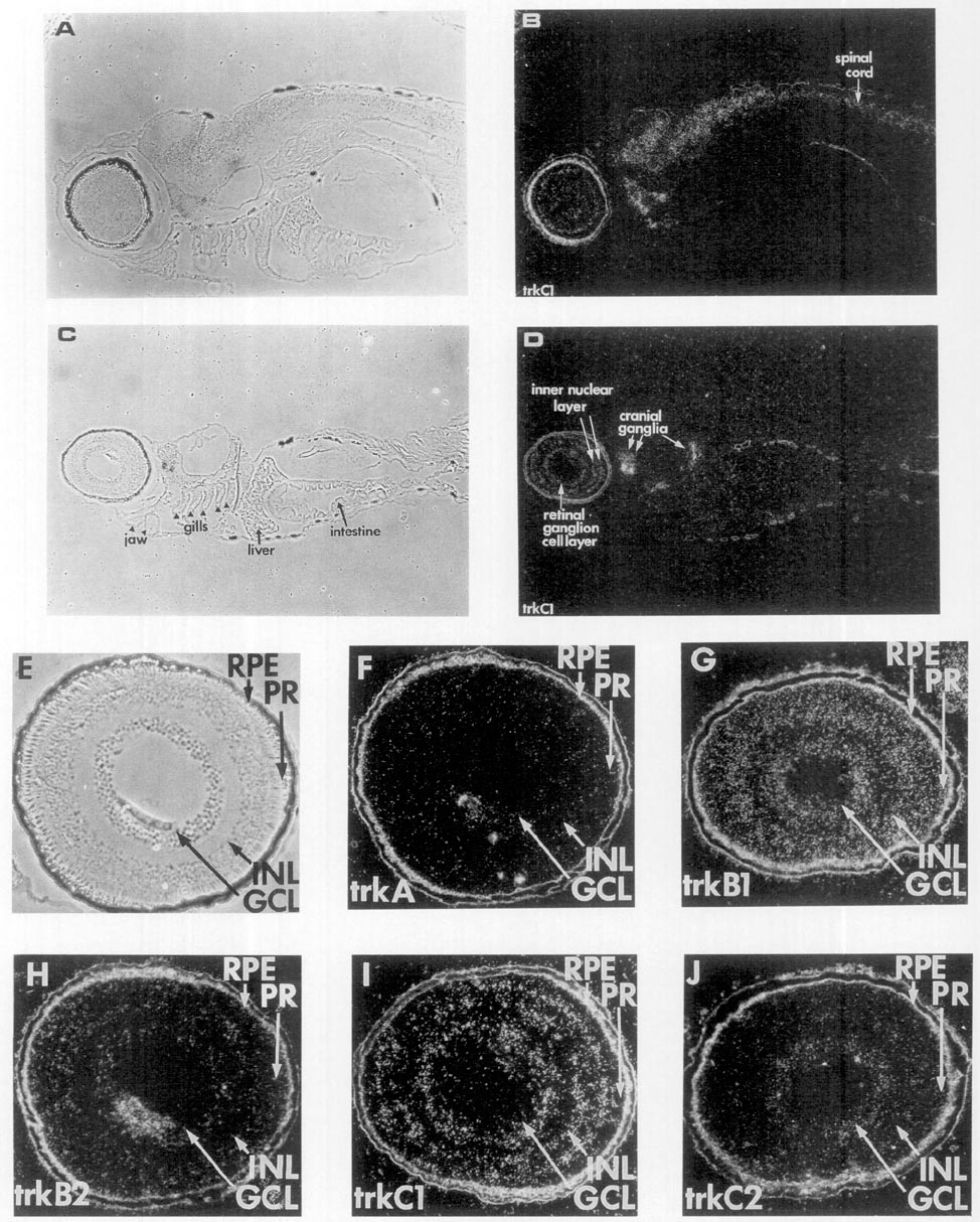
Fig. 7 Zebrafish trk expression in the spinal cord, cranial ganglia, and retina. RNA probes labeled with [35S]UTP specific for trk full-length transcripts were used to examine expression 6 days after fertilization, in sagittal sections for trkC1 (A-D) and the retina for the five trk probes (E-J) using in situ hybridization. (B and D) trkC1 dark-field photomicrographs. (A and C) phase photomicrographs of B and D, respectively. (F) trkA, (G) trkB1, (H) trkB2, (I) trkC1, and (J) trkC2 are dark-field photomicrographs. (E) Phase photomicrograph of F (trkA). The positions of retinal pigment epithelium (RPE), photoreceptors (PR), inner nuclear layer (INL), and ganglion cell layer (GCL) are indicated. The lens next to the GCL in F and H appears white but does not correspond to labeling. The scale bar is 50 μm.
Reprinted from Developmental Biology, 169, Martin, S.C., Marazzi, G., Sandell, J.H., and Heinrich, G., Five Trk receptors in the zebrafish, 745-758, Copyright (1995) with permission from Elsevier. Full text @ Dev. Biol.