
- Title
-
Holoprosencephaly and cyclopia in bmp7b and bmpr1ba Crispant zebrafish
- Authors
- Kyrychenko, V., Rensinghoff, P., Bulk, J., Frey, C., Heermann, S.
- Source
- Full text @ Animal Cells Syst (Seoul)
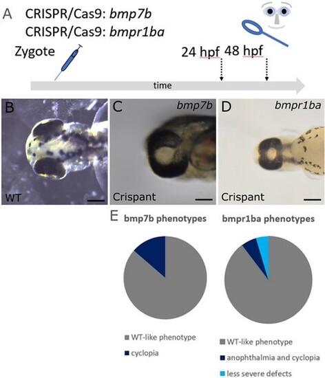
Targeting of bmp7b and bmpr1ba with CRISPR/Cas9 results in cyclopia. (A) Scheme of experimental procedure. Embryos were injected at 1-cell stage and analyzed at approximately 24–48 hpf. (B) Wild-type, dorsal view. (C) bmp7b Crispant with cyclopia, ventral view. (D) bmpr1ba Crispant with cyclopia, ventral view. Scalebars indicate 200 µm. (E) Pie charts show the frequency of phenotypes in surviving embryos in bmp7b and bmpr1ba Crispants. PHENOTYPE:
|
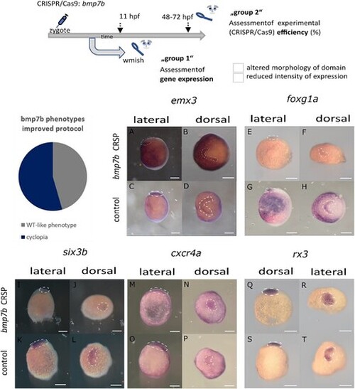
Analysis of gene expression patterns (wmish) in ANP domains in Crispants for bmp7b and controls (I) (embryos are presented in a lateral and in a dorsal view, the presumptive head is facing to the left, respectively, in dorsal views, expression domains are encircled). Scheme (top) showing the experimental procedure: CRISPR/Cas9 injections into zygotes, subsequent separation of embryos: group 1: wmish analysis, group 2: assessment of experimental efficiency (please see the Table; 5–6 uninjected embryos served as controls for each experiment). Three experiments were evaluated for the analysis of individual genes. Binary values were assigned to the Crispants: ‘normal’ vs. ‘altered’ with respect to ‘altered morphology of domain’ and ‘reduced intensity of expression’, respectively. Results are given in the Table. Pie chart shows the frequency of phenotypes in surviving embryos with the improved protocol. (A–D) show expression patterns of emx3 in controls (C, D) and Crispants (A, B). Please note the reduction of expression in the lateral domain (right) in (B). (E–H) show expression of foxg1a in controls (G, H) and Crispants (E, F). Please note the reduction of expression most seen in the lateral domain (right) in (F). (I–L) show expression of six3b in controls (K, L) and Crispants (I, J). Please note the condensed expression domain closer to the midline (J). (M–P) show expression of cxcr4a in controls (O, P) and Crispants (M, N). Please note the condensed expression domain closer to the midline (N). (Q–T) show expression of rx3 in controls (S, T) and Crispants (Q, R). Please note the condensed expression domain closer to the midline (R). EXPRESSION / LABELING:
PHENOTYPE:
|
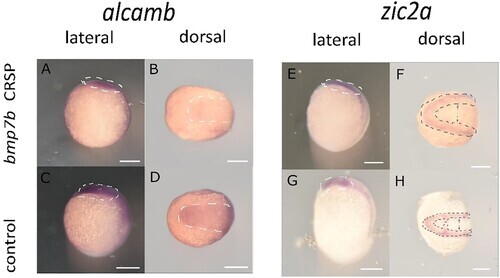
Analysis of gene expression patterns (wmish) in ANP domains in Crispants for bmp7b and controls (II). (embryos are presented in a lateral and in a dorsal view, the presumptive head is facing to the left, expression domains are encircled). The experimental design is as presented in Figure 3. (A–D) show expression of alcamb in controls (C, D) and Crispants (A, B). Please note the reduced expression in B in comparison to C (encircled). (E–H) show expression of zic2a in controls (G, H) and Crispants (E, F). Please note the extended width in between the lateral expression domains (arrows in F in comparison to arrows in H) as well as the reduced expression at the prospective posterior domain of the ANP (encircled in H, almost absent in F). EXPRESSION / LABELING:
PHENOTYPE:
|
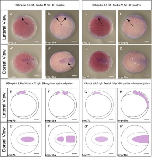
Expression patterns and domains of bmp7b and bmpr1ba in inner-embryonic domains and structures after induction of bmp4 and in controls (lateral and dorsal views, the prospective head facing left). Scalebars indicate 200 µm. Scheme of experimental procedure (top): Heat shock of outcrossed transgenes tg(hsp:bmp4/cmlc2:GFP) at 8.5 hpf and fixation for whmish at 11 hpf. Genotyping and separation of recorded expression patterns (A–B′) specimens with typical expression patterns, genotyped cmlc2:GFP/bleeding heart (BH) negative. (C–D′) Specimens with typical expression patterns genotyped cmlc2:GFP/BH positive. (E–H′) Schematic drawing of the respective patterns. (A, A′, E and E′) Expression pattern of bmp7b without bmp4 induction (3 embryos, 3 embryos with pattern). Note the broad expression domain, at the anterior midline, likely corresponding to the anterior neuroectoderm. (B, B′, F and F′) Expression domain of the bmpr1ba without bmp4 induction (5 embryos, 5 embryos with pattern). Please note the two distinct and separate expression domains. The anterior domain, likely corresponds to the anterior ANP. The posterior domain likely corresponds to the region of the midbrain-hindbrain boundary. (C, C′, G and G′) Expression domain of bmp7b after induction of bmp4 (8 embryos, 6 embryos with pattern). The expression domain remains similar to the uninduced one, albeit with a slight smaller projection dorsally, seen in the lateral perspective (please note the arrow in (C)). (D, D′, H and H′) Expression domain of bmpr1ba after induction of bmp4 (16 embryos, 16 embryos with pattern). Please note the single expression domain, which mostly includes the domains seen in the control. No gap is seen separating an anterior and a posterior expression domain as seen in the control. The anterior margin of the ANP lacks expression (arrow in (D)), please see (B) as control. EXPRESSION / LABELING:
PHENOTYPE:
|
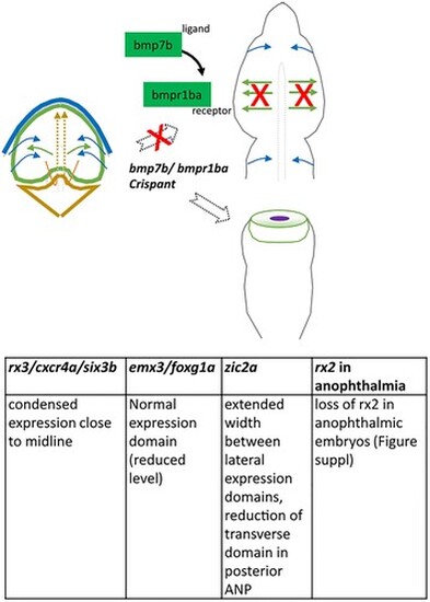
Schematic and tabular summary of major findings: In Crispants for bmp7b (and bmpr1ba) the separation of the eye field fails during early development and the development results in cyclopia. Genes expressed in the eye field rx3, cxcr4a and six3b are condensed to the midline in Crispants (shown for bmp7b). The morphology of the zic2a expression domain of was also found altered in Crispants. Zic2 was found to be important for prechordal plate development and is thus a HPE-related gene (Warr et al. Citation2008). In Crispants, however, the domain was showing an extended width, while the domain marking the posterior end of the ANP was largely missing. This domain is corresponding to the presumptive hypothalamic domain, which is important for ANP splitting via a subduction movement (England et al. Citation2006). Alcamb (formerly nlcam), which in Medaka was found to be suppressed in retinal precursors (Brown et al. Citation2010), was found homogeneously expressed in the ANP of control zebrafish embryos and reduced in Crispants. Genes expressed in the prospective telencephalic domain, emx3 and foxg1a, show an overall normal morphology of their expression domain (in terms of separation from the midline) but, however, reduced levels of expression intensity. In cases of Crispants resulting in anophthalmia, no ‘crypt-oculoid’ was found. |