FIGURE SUMMARY
Title

Antioxidant, Osteogenic, and Neuroprotective Effects of Homotaurine in Aging and Parkinson's Disease Models

Authors
Minoia, A., Piritore, F.C., Bolognin, S., Pessoa, J., Bernardes de Jesus, B., Tiso, N., Romanelli, M.G., Schwamborn, J.C., Dalle Carbonare, L., Valenti, M.T.
Source
Full text @ Antioxidants (Basel)

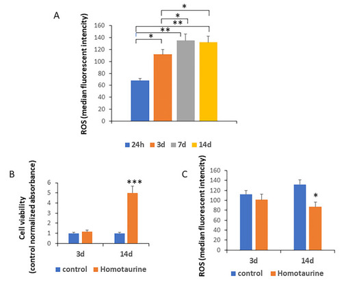
Assessment of ROS levels, MSC viability, and the effect of homotaurine treatment during a 14-day period. (A) A significant increase in reactive oxygen species (ROS) levels was observed starting from day 3, suggesting a correlation between elevated ROS and cellular aging in mesenchymal stem cells (MSCs). (B) No significant differences in cell viability were noted after 3 days of culture, while a marked improvement in cell viability was observed on day 14. (C) This cell viability increase was associated with a reduction in ROS levels following homotaurine treatment, also on day 14, with no previous alteration after 3 days of treatment. (* p < 0.05; ** p < 0.01; *** p < 0.005).

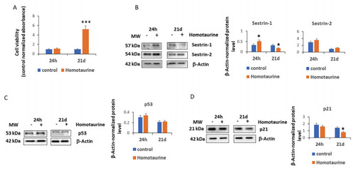
Effect of homotaurine on cell viability, sestrin expression, and cell cycle regulators in MSCs at 24 h and 21 days of treatment. (A) The positive effects of homotaurine on cell viability were observed after 21 days of treatment. (B) A significant increase in sestrin 1 levels was detected after 24 h of treatment; however, a decrease was detected at 21 days of homotaurine treatment, while sestrin 2 levels remained unchanged. (C) p53 levels did not show any significant changes in homotaurine-treated cells, while (D) a significant reduction in p21 levels was observed after 21 days of treatment. (* p < 0.05; *** p < 0.005). The original Western blot images are provided in the Supplementary Materials S1 and S2.

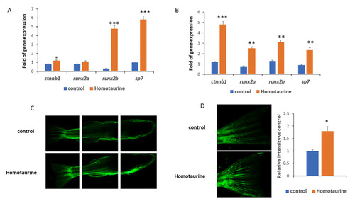
Effects of homotaurine on osteogenesis and angiogenesis gene expression in zebrafish. (A) In zebrafish larvae treated for 14 days, homotaurine significantly increased the expressions of the ctnnb1, runx2b, and sp7 genes, crucial for skeletal formation. (B) Similar effects on the skeletal gene expression levels of ctnnb1, runx2b, runx2b, and sp7 were observed in 4-month-old zebrafish. (C,D) In aged zebrafish (36 months old) treated with homotaurine for 7 days, (C) vascular endothelial growth factor receptor 2 (VEGFR2) expression was notably enhanced, (D) particularly in the tail region, as visualized in the kinase insert domain receptor-like–enhanced green fluorescent protein (KDRL-EGFP) transgenic model. Magnifications 4x (C) and 10x (D); (* p < 0.05; ** p < 0.01; *** p < 0.005).

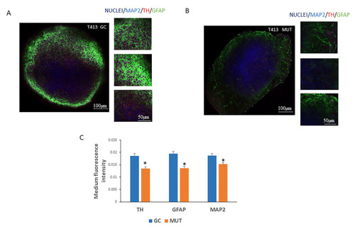
Expression of neuronal markers in 3D neuronal epithelial stem cell (NESC) organoids derived from induced pluripotent stem cells (iPSCs) carrying the leucine-rich repeat kinase 2 (LRRK2) G2019S mutation. (A) Three-dimensional NESC organoids generated from both gene-corrected (GC) and (B) mutated (MUT T413) iPSC lines were stained with antibodies against TH, GFAP, and MAP2 neuronal markers and visualized using confocal microscopy. Representative micrographs are shown. (C) Through a semi-quantitative analysis of the images, we observed reductions in the expression levels of all examined neuronal markers: tyrosine hydroxylase (TH), glial fibrillary acidic protein (GFAP), and microtubule-associated protein 2 (MAP2) in the NESC 3D colonies derived from the LRRK2-G2019S-mutated T413 cell line, compared to the gene-corrected GC counterpart. In blue nuclei, in purple the MAP2 marker, in red the TH marker and in green the GFAP marker (* p < 0.05).

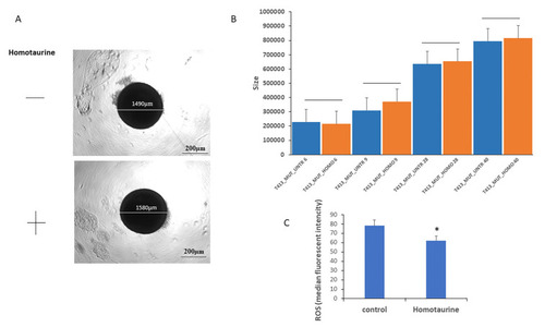
Evaluation of homotaurine’s effects on LRRK2-G2019S mutated midbrain organoids size and ROS levels during differentiation in 8 pooled organoids for each condition. (A) Representative images of mock-treated (top) and homotaurine-treated (bottom) NESC colonies, whose diameters are indicated. (B) The area (size, µm2) of LRRK2-G2019S-mutated midbrain organoids derived from mutated iPSC lines was assessed at multiple differentiation time points (6, 9, 28, and 40 days). Homotaurine treatment did not alter colony size compared to untreated (UNTR) controls, indicating that homotaurine does not adversely affect colony growth during differentiation. (C) Cytofluorimetric analysis revealed a significant reduction in ROS levels in homotaurine-treated samples at day 40 of differentiation. (* p < 0.05).

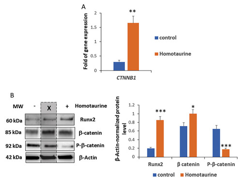
Modulation of Wnt signaling effectors by homotaurine treatment of LRRK2-G2019S-mutated midbrain organoids. (A) Gene expression analysis for 4 pooled organoids revealed an increase in CTNNB1 gene expression levels in response to homotaurine treatment. (B) Protein analysis for 4 pooled organoids showed an increase in total β-catenin and RUNX2, along with a decrease in the phosphorylated form of β-catenin, indicating the activation of the Wnt signaling pathway. (* p < 0.05; ** p < 0.01; *** p < 0.005). In the Western blot images, the lanes in the middle marked with the ‘X’ do not contain samples from this study. The original Western blot images are provided in Supplementary Materials S3.

Acknowledgments
This image is the copyrighted work of the attributed author or publisher, and ZFIN has permission only to display this image to its users. Additional permissions should be obtained from the applicable author or publisher of the image. Full text @ Antioxidants (Basel)