FIGURE SUMMARY
Title

New insights into the all-testis differentiation in zebrafish with compromised endogenous androgen and estrogen synthesis

Authors
Ruan, Y., Li, X., Wang, X., Zhai, G., Lou, Q., Jin, X., He, J., Mei, J., Xiao, W., Gui, J., Yin, Z.
Source
Full text @ PLoS Genet.

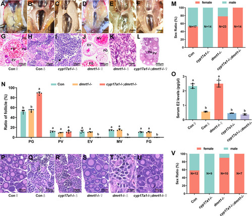
Additional depletion of dmrt1 restored the phenotype of ovary differentiation in cyp17a1-/- zebrafish.

(A–F) Anatomical examination of the gonads from the control fish, cyp17a1-/- fish, dmrt1-/- fish, and cyp17a1-/-;dmrt1-/- fish at 90 dpf. (G–L) Histological analysis of the gonads from the control fish, cyp17a1-/- fish, dmrt1-/- fish, and cyp17a1-/-;dmrt1-/- fish at 90 dpf. (A and G) Control fish ovary. (M) Sex ratios in fish of each genotype mentioned above at 90 dpf. (N) Ratios of PG, PV, EV, MV and FG follicles in fish of each genotype at 90 dpf. PG, primary growth. PV, previtellogenic. EV, early vitellogenic. MV, middle vitellogenic. FG, full grown. (O) Concentration of serum estradiol in control females, control males, cyp17a1-/- fish, dmrt1-/- females, and cyp17a1-/-;dmrt1-/- fish. E2, estradiol. (P–U) Histological analysis of the gonads from the control fish, cyp17a1-/- fish, dmrt1-/- fish, and cyp17a1-/-;dmrt1-/- fish at 50 dpf. (V) Sex ratios in fish of each genotype mentioned above at 50 dpf. Different letters in the bar charts represent significant differences.

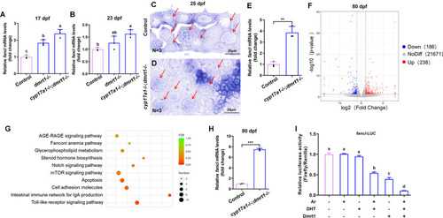
The cyp17a1-/-;dmrt1-/- fish exhibited increased expression of fancl.

(A and B) Relative expression of fancl in control fish, dmrt1-/- fish and cyp17a1-/-;dmrt1-/- fish at 17 dpf and 23 dpf was tested with qPCR. For fish RNA sampling at 17 dpf and 23 dpf, every 5 body trunks of fish collected were mixed into one sample, and 3 samples were examined. (C and D) In situ hybridization was performed on cryosections of presumptive ovaries from control fish and cyp17a1-/-;dmrt1-/- fish at 25 dpf using the antisense probe of fancl. Arrows point to the immature oocytes. (E) Comparison of differentially expressed gene in the ovaries of control fish and cyp17a1-/-;dmrt1-/- fish at 80 dpf. Volcano plot shows genes that were differentially expressed in ovaries between control and cyp17a1-/-;dmrt1-/- fish. (F) Gene set enrichment analysis based on genes differentiated expressed in cyp17a1-/-;dmrt1-/- fish at 80 dpf. The length of the bar represents the false discovery rate (FDR). (G) Relative expression of fancl in the ovaries of control fish and cyp17a1-/-;dmrt1-/- fish at 80 dpf with qPCR. For fish at 80 dpf, every three dissected ovaries were mixed as one sample, and 3 samples were examined. (H) The effect of Dmrt1 and DHT/Ar in regulating the relative luciferase activity driven by fancl promoter. DHT, dihydrotestosterone. ***, p < 0.001. Different letters in the bar chart represent significant differences.

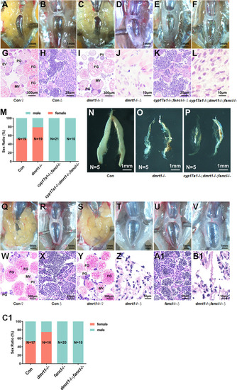
Additional depletion of fancl blocked female-biased sex ratio of cyp17a1-/-;dmrt1-/- fish and dmrt1-/- fish.

(A–F) Anatomical examination of the gonads from the control fish, dmrt1-/- fish, cyp17a1-/-;fancl-/- fish, and cyp17a1-/-;dmrt1-/-;fancl-/- fish. (G–L) Histological analysis of the gonads from the control fish, dmrt1-/- fish, cyp17a1-/-;fancl-/- fish and cyp17a1-/-;dmrt1-/-;fancl-/- fish. (M) Sex ratios in fish of each genotype mentioned above at 90 dpf. (N-P) The visualization of dissected testis of control fish, dmrt1-/- fish and cyp17a1-/-;dmrt1-/-;fancl-/- fish. (Q–V) Anatomical examination of the gonads from the control fish, dmrt1-/- fish, fancl-/- fish, and dmrt1-/-; fancl-/- fish. (W–B1) Histological analysis of the gonads from the control fish, dmrt1-/- fish, fancl-/- fish, and dmrt1-/-; fancl-/- fish. (C1) Sex ratios in fish of each genotype mentioned above at 90 dpf. PG, primary growth. PV, previtellogenic. EV, early vitellogenic. MV, middle vitellogenic. FG, full growth.

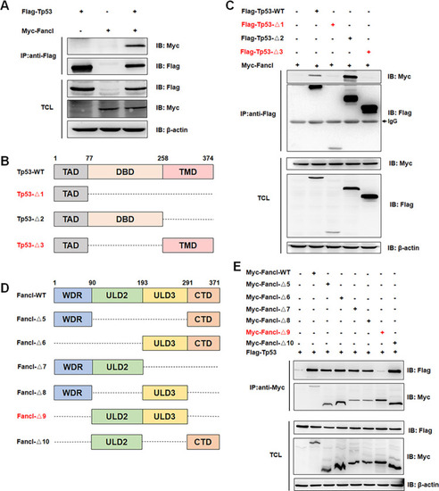
Fancl interacts with Tp53 in HEK293T cells.

(A) The interaction of Fancl with Tp53 in HEK293T cells as revealed by the Co-IP assay. Myc-tagged Fancl and Flag-tagged Tp53 were transfected into HEK293T cells, then anti-Flag antibody-conjugated agarose beads were used for immune-precipitation. (B and C) Domain mapping revealed that the DBD domain of Tp53 is required for their interaction. TAD, transactivation domain. DBD, DNA binding domain. TMD, tetramerization domain. (D and E) Domain mapping revealed that the multiple domains, WDR and CTD, of Fancl are required for their interaction. WDR, WD-repeat domain. ULD2, UBC-like domain 2. ULD3, UBC-like domain 3. CTD, C-terminal domain. IP, immunoprecipitation. IB, immunoblotting. TCL, total cell lysate.

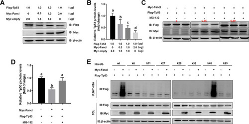
Fancl promoted K48-linked ubiquitination of Tp53 in HEK293T cells.

(A) Transfection of Myc-tagged Fancl decreased the levels of Flag-tagged Tp53 in a dose-dependent manner. (B) Quantification of the western blot bands of the target protein, Tp53. (C) The proteasome inhibitor, MG-132, blocked the Fancl-mediated Tp53 destabilization. (D) Quantification of the western blot bands of the target protein, Tp53. (E) Fancl promotes K48-linked ubiquitination, rather than K6-, K11-, K27-, K29-, K33-, K63-linked ubiquitination of Tp53. IP, immunoprecipitation. IB, immunoblotting. TCL, total cell lysate. Different letters in the bar charts represent significant differences.

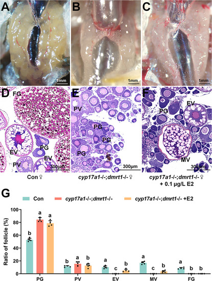
Administration of 17β-estradiol rescued the arrested folliculogenesis of cyp17a1-/-;dmrt1-/- fish.

(A–C) Histological analysis of the ovaries from the control fish, cyp17a1-/-;dmrt1-/- fish, and cyp17a1-/-;dmrt1-/- fish administrated 17β-estradiol from 80 to 110 dpf. (D–F) Anatomical examination of the ovaries from the control fish, cyp17a1-/-;dmrt1-/- fish and cyp17a1-/-;dmrt1-/- fish administrated 17β-estradiol from 80 to 110 dpf. (G) Ratios of PG, PV, EV, MV and FG follicles in fish of each genotype at 110 dpf. PG, primary growth. PV, previtellogenic. EV, early vitellogenic. MV, middle vitellogenic. FG, full grown. Different letters in the bar chart represent significant differences.

Acknowledgments
This image is the copyrighted work of the attributed author or publisher, and ZFIN has permission only to display this image to its users. Additional permissions should be obtained from the applicable author or publisher of the image. Full text @ PLoS Genet.