IMAGE

Fig 1

ID
ZDB-IMAGE-240310-2
Source
Figures for Ruan et al., 2024
Image
Figure Caption

Fig 1 Additional depletion of dmrt1 restored the phenotype of ovary differentiation in cyp17a1-/- zebrafish.

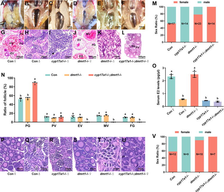
(A–F) Anatomical examination of the gonads from the control fish, cyp17a1-/- fish, dmrt1-/- fish, and cyp17a1-/-;dmrt1-/- fish at 90 dpf. (G–L) Histological analysis of the gonads from the control fish, cyp17a1-/- fish, dmrt1-/- fish, and cyp17a1-/-;dmrt1-/- fish at 90 dpf. (A and G) Control fish ovary. (M) Sex ratios in fish of each genotype mentioned above at 90 dpf. (N) Ratios of PG, PV, EV, MV and FG follicles in fish of each genotype at 90 dpf. PG, primary growth. PV, previtellogenic. EV, early vitellogenic. MV, middle vitellogenic. FG, full grown. (O) Concentration of serum estradiol in control females, control males, cyp17a1-/- fish, dmrt1-/- females, and cyp17a1-/-;dmrt1-/- fish. E2, estradiol. (P–U) Histological analysis of the gonads from the control fish, cyp17a1-/- fish, dmrt1-/- fish, and cyp17a1-/-;dmrt1-/- fish at 50 dpf. (V) Sex ratios in fish of each genotype mentioned above at 50 dpf. Different letters in the bar charts represent significant differences.

Acknowledgments
This image is the copyrighted work of the attributed author or publisher, and ZFIN has permission only to display this image to its users. Additional permissions should be obtained from the applicable author or publisher of the image. Full text @ PLoS Genet.